A trio of researchers at MRC Laboratory of Molecular Biology has developed a support film for creating sharper images in cryo-electron microscopy. In their paper published in the journal Science, Katerina Naydenova, Peipei Jia and Christopher Russo describe the factors that lead to blurring due to sample movement. They also describe the support film they developed to correct the problem. Micah Rapp and Bridget Carragher with Columbia University have published a Perspective piece in the same journal issue describing the work by the team and how the new support film is likely to impact future research efforts.
Cryogenic electron microscopy developed to the point of being useful in the 1970s, and since that time, the technique for studying materials at near-atomic resolution has steadily improved—so much, in fact, that advances that once took decades can now be achieved in months. The speedy understanding of the SARS-CoV-2 virus structure is one recent example. In this new effort, the researchers have found a way to improve the resolution of images even further by addressing one of the inherent limitations of the technique—movement of the sample during scanning, which leads to blurring.
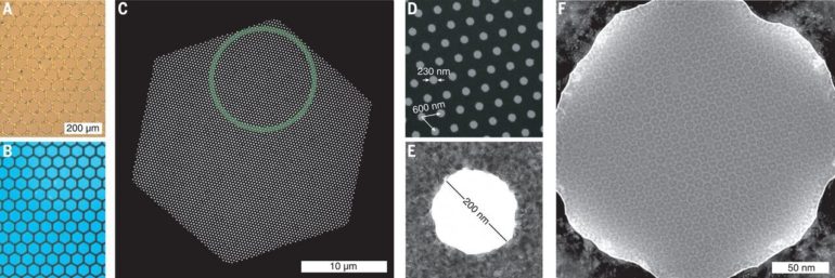
Cryogenic electron microscopy involves placing material to be sampled in water droplet, which is then applied to a grid mesh. Once in place, the mesh with its sample is dunked in liquid ethane to make it extremely cold. At that point, the sample is ready for study, though it is still susceptible to slight movements during the scanning process. The researchers began their work by studying samples to better understand why they moved during scanning. They discovered that the primary reason was vitrified ice layer buckling. After some tinkering, they found they could reduce buckling by optimizing the thickness of the grid by using gold and making the holes in the mesh more densely arranged. They named the new support film “hexAuFoil.” Testing showed greatly reduced buckling, which in turn greatly reduced the movement of samples, resulting in shaper sample images. The researchers found that their new support film reveals features in samples that until now had been lost during scanning.
New microscopy technique improves imaging at the atomic scale
More information:
K. Naydenova el al., Cryo–electron microscopy with sub-angstrom specimen movement, Science (2020). science.sciencemag.org/cgi/doi … 1126/science.abb7927
M. Rapp el al., Better, faster, and even cheap, Science (2020). science.sciencemag.org/cgi/doi … 1126/science.abd8035
2020 Science X Network
Citation:
Support film makes cryo-electron microscopy sharper (2020, October 9)
retrieved 11 October 2020
from https://phys.org/news/2020-10-cryo-electron-microscopy-sharper.html
This document is subject to copyright. Apart from any fair dealing for the purpose of private study or research, no
part may be reproduced without the written permission. The content is provided for information purposes only.