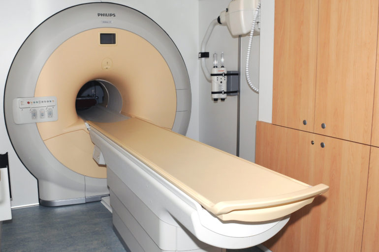
Increased immune system activity along the surface of the brain, or meningeal inflammation, may be important for understanding how multiple sclerosis (MS) progresses from the most common and earliest form of the disease known as relapsing remitting MS (RRMS) to a secondary progressive form. The meninges are a thin, protective tissue that covers the brain and spinal cord. One proposed way to more easily see evidence of inflammation at the meninges is by finding leptomeningeal enhancement (LME) on Magnetic Resonance Imaging (MRI) scans. This can appear early in the course of the disease and increases as the disease progresses, yet the most commonly available MRI technology—3 Tesla (3T)—gives a limited view of this proposed marker. Investigators from Brigham and Women’s Hospital have completed a new study using 7 Tesla (7T) MRI—a far more powerful imaging technology—to further examine LME in MS patients. With this new powerful tool, they have found that this proposed marker of brain inflammation in MS patients is more common than previously reported and is tied to lesions in the gray matter regions of the brain. The team’s findings are published in the Multiple Sclerosis Journal.
“The 7T MRI scanner affords us new ways of viewing areas of damage in neurologic diseases such as MS that were not well seen using 3T MRI; it’s capturing nuances that we would otherwise miss,” said corresponding author Jonathan Zurawski, MD, a neurologist at the Brigham. “The 7T scanner reveals markers or signatures that were poorly…

