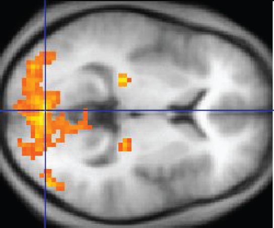
Research led by a Wayne State University Department of Mathematics professor is aiding researchers in Wayne State’s Department of Psychiatry and Behavioral Neurosciences in analyzing fMRI data. fMRI is the preeminent class of signals collected from the brain in vivo and is irreplaceable in the study of brain dysfunction in many medical fields, including psychiatry, neurology and pediatrics.
Andrew Salch, Ph.D., associate professor of mathematics in Wayne State’s College of Liberal Arts and Sciences, is leading the multidisciplinary team that is investigating how concepts of topological data analysis, a subfield of mathematics, can be applied to recovering “hidden” structure in fMRI data.
“We hypothesized that aspects of the fMRI signal are not easily discoverable using many of the standard tools used for fMRI data analysis, which strategically reduce the number of dimensions in the data to be considered. Consequently, these aspects might be uncovered using concepts from the mathematical field of topological data analysis, also called TDA, which is intended for use on high-dimensional data sets,” said Salch. “The high dimensionality that characterizes fMRI data includes the three dimensions of space—that is, where in the brain the signal is being acquired—time—or how the signal varies as brain states change in time—and signal intensity—or how the strength of the fMRI signal changes in response to the task. When related to task-induced changes, the results reflect biologically meaningful aspects of brain function and dysfunction. This is a unique collaborative work focused on the complexities of both TDA and fMRI respectively, show how TDA can be applied to real fMRI data collected, and provide open access computational software we have developed for implementing the analyses.”
The research article, “From mathematics to medicine: A practical primer on topological data analysis and the development of related analytic tools for the functional discovery of latent structure in fMRI data,” appears in the Aug. 12 issue of PLOS ONE.
In it, the team used TDA to discover data structures in the anterior cingulate cortex, a critical control region in the brain. These structures—called non-contractible loops in TDA—appeared in specific conditions of the experiment, and were not identified using conventional techniques for fMRI analyses.
“We expect this work to become a citation classic,” said Vaibhav Diwadkar, Ph.D., professor of psychiatry and behavioral neurosciences and research collaborator. “Instead of merely applying TDA to fMRI, we provide a lucid argument for why medical researchers who use fMRI should consider using TDA, and why topologists should turn their attention to the study of complex fMRI data. Moreover, this important work provides readers with empirical demonstrations of such applications, and we provide potential users with the tools we used so they can in turn apply it to their own data.”
“Our ongoing research utilizing TDA with fMRI will provide a unique and complementary method for assessing brain function, and will give medical researchers greater flexibility in tackling complex properties in their data,” said Salch. “In particular, our work will help fMRI researchers become aware of the significant power of TDA that is designed to address complexity in data, and will enhance the value of using fMRI in neuroscience and medicine.”
New approach simultaneously measures EEG and fMRI connectomes
More information:
Andrew Salch et al, From mathematics to medicine: A practical primer on topological data analysis (TDA) and the development of related analytic tools for the functional discovery of latent structure in fMRI data, PLOS ONE (2021). DOI: 10.1371/journal.pone.0255859
Provided by
Wayne State University
Citation:
From mathematics to medicine: Applying complex mathematics to analyze fMRI data (2021, August 18)
retrieved 18 August 2021
from https://medicalxpress.com/news/2021-08-mathematics-medicine-complex-fmri.html
This document is subject to copyright. Apart from any fair dealing for the purpose of private study or research, no
part may be reproduced without the written permission. The content is provided for information purposes only.


