by KeAi Communications Co.
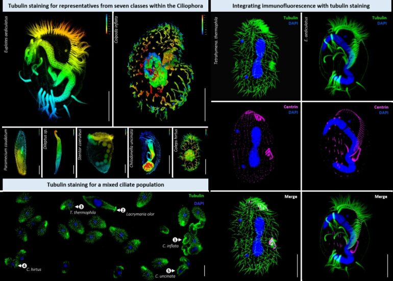
Ciliates, a group of single-celled ciliated eukaryotes, have been studied since the dawn of light microscopy, with over 10,000 species described. Cilia are the key feature of ciliates and contribute to their diversity. They vary in length, covering the cell surface or specific parts, or can bundle to form cirri. Staining cilia and basal bodies is a common procedure in ciliate morphology research, using techniques like silver impregnation and immunofluorescence staining with antibodies associated with tubulin.
Live-cell tubulin dyes, commonly used in human cells, require cell permeabilization reagents to enter the cells. However, these reagents are ineffective in ciliates. In ciliate cells, the dyes are usually ingested through phagocytosis and end up in membrane-enclosed organelles, posing a challenge in delivering the dyes.
In a study published in the journal Water Biology and Security, a team of researchers from China developed a straightforward and adaptable method for tubulin/cilia staining that yields excellent results in ciliates.
“Our method entails utilizing live-cell tubulin dyes to stain fixed ciliate cells, presenting a highly efficient and practical option for ciliate research,” says lead author Tingting Pan. “Moreover, it allows for seamless integration immunofluorescence staining with antibodies when required.”
This new tubulin-staining method offers the ciliate research community four significant advantages.
It boosts cell biology studies in ciliates, particularly in cytoskeleton research, as tubulin cytoskeletons are prevalent in these organisms
It simplifies the staining process, benefiting morphology and taxonomy studies and making it accessible to most ciliate taxonomy laboratories
When combined with confocal microscopy, the method allows for detailed exploration of basal body patterns—crucial taxonomic features—using only a few cells and bypassing the complexities of traditional silver impregnation methods
Ecological studies of ciliates stand to benefit from this method.
“The method flexibility enables the staining of mixed cell populations from aquatic environments, post-enrichment using a plankton net,” adds Pan. “As high-throughput fluorescence imaging advances, our staining method could seamlessly integrate with machine learning-based classification systems due to its capacity for images with clear backgrounds.”

More information:
Tingting Pan et al, An improved method for tubulin staining of ciliated eukaryotes, Water Biology and Security (2024). DOI: 10.1016/j.watbs.2024.100274
Provided by
KeAi Communications Co.
Citation:
Ciliated eukaryotes study offers simple, versatile method for tubulin staining (2024, July 22)

