Animals showcase a remarkable diversity of colors and patterns, from the shimmery appearance of a peacock’s tail to the distinctive rosettes on a jaguar’s fur. Quantifying animal color has been a longtime goal of evolutionary biologists, who aim to understand how color evolved over time—and the physical and genetic mechanisms involved.
Ultimately, studying animal color is important because it can reveal how evolutionary forces, such as natural and sexual selection, favor certain traits over others. However, fully capturing animal color is challenging because researchers must choose between high spatial resolution (as in traditional photography, which captures information in a limited number of color channels) and high spectral resolution (as in spectrophotometry, which captures a reflectance spectrum at a single point).
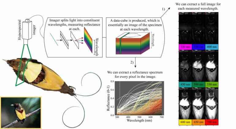
Evolutionary biologists at Princeton University recently used hyperspectral imaging, a state-of-the-art tool that measures detailed spectral information at each pixel in an image, to investigate avian plumage color. Hyperspectral imaging works by separating the light spectrum into a series of narrow bands, each corresponding to a small range of wavelengths.
Essentially, an image is taken in each of these narrow bands, generating a stack of images (or a “data-cube”) that includes both spatial and spectral information. Each pixel in the data-cube contains detailed information about the wavelengths of light reflected.
“Hyperspectral imaging offers the best of both worlds,” explained Dr. Mary Caswell Stoddard, a professor in the Department of Ecology and Evolutionary Biology and the study’s senior author. “Researchers can capture comprehensive reflectance data for an entire specimen in a matter of minutes, opening up new possibilities for the study of animal color.”
Hyperspectral imaging, often used in agricultural and medical applications, has been used in a handful of studies on animal color, but uptake has generally been slow. Hyperspectral data can be unwieldy—and commercial cameras are expensive and rarely capture all of the wavelengths relevant to animals.
“In our study, we developed a new computational pipeline—a series of step-by-step analyses—to show how researchers can obtain and study hyperspectral data from museum specimens. We published all the hyperspectral data we collected, as well as all of the code we developed, to aid others in replicating and building on our methods,” said Dr. Ben Hogan, an associate research scholar and the study’s lead author.
Hogan and Stoddard used a commercial camera sensitive to wavelengths ranging from 325 to 700 nanometers, which largely corresponds to the spectrum visible to birds (typically 300 to 700 nanometers), including the ultraviolet range (300 to 400 nanometers).
Many birds have feathers that reflect ultraviolet light. “Using hyperspectral imaging, we can easily capture detailed ultraviolet images, sometimes revealing entire patches of ultraviolet color that are invisible to humans,” said Stoddard.
To demonstrate the power of hyperspectral imaging in animal color research, Hogan and Stoddard focused on the birds-of-paradise. These charismatic birds are native to New Guinea and nearby regions and are known for their vibrant plumage and elaborate courtship displays.
Discover the latest in science, tech, and space with over 100,000 subscribers who rely on Phys.org for daily insights.
Sign up for our free newsletter and get updates on breakthroughs,
innovations, and research that matter—daily or weekly.
The rare hybrid King of Holland’s bird-of-paradise was the key subject of interest. Only about 25 male museum specimens are known to exist worldwide, with 12 specimens held by the American Museum of Natural History (AMNH) in New York City. The hybrid, a cross between the King and Magnificent birds-of-paradise, has plumage that appears to combine features of its two parent species.
By collecting and analyzing hyperspectral data from specimens borrowed from the AMNH, Hogan and Stoddard were able to quantify the degree to which the hybrid’s appearance was truly intermediate—that is, a precise blend of colors from its parent species.
“We were surprised to discover that for several plumage patches—even those colored by very specific micro- and nano-structures—the hybrid’s color really does resemble a mixture of those from the parental phenotypes,” said Hogan.
Hogan and Stoddard also integrated hyperspectral imaging with photogrammetry, a technique that stitches together hundreds of traditional images taken from different angles, to produce virtual 3D models of the bird specimens. These 3D models are valuable because they reveal how an animal’s body shape and morphology interact with its coloration. They also provide detailed digital records of specimens, which can be easily accessed by researchers and the public—and used in a variety of morphometric analyses.
Hyperspectral imaging will be a powerful tool for studying camouflage, warning coloration, mimicry and courtship displays in birds—and beyond. The technique is ideal for investigating other colorful taxonomic groups, such as butterflies and beetles. In the future, 3D models integrated with hyperspectral data could be animated to explore how motion influences signal design.
“We imagine that hyperspectral imaging, combined with 3D modeling, could become the new ‘gold standard’ for many studies of animal coloration, particularly those based in museum collections,” said Stoddard. “Although hyperspectral imaging of moving animals in the field remains a challenge—as does capturing iridescent color—the approach has tremendous potential.”
The study is published in PLOS Biology.
More information:
Benedict G. Hogan et al, Hyperspectral imaging in animal coloration research: A user-friendly pipeline for image generation, analysis, and integration with 3D modeling, PLOS Biology (2024). DOI: 10.1371/journal.pbio.3002867
Provided by
Princeton University
Citation:
Hyperspectral imaging technique illuminates the colorful plumage of birds (2024, December 9)


