Researchers at University of Tsukuba have developed a method for culturing fertilized chick eggs without their shells. The eggs were placed in an artificial culture vessel made of transparent film, allowing for real-time observation of the chick embryo’s development from laying to hatching.
The study is published in the journal Scientific Reports.
Efforts to observe chicken embryonic development date back to Aristotle (300 BC). However, because the chicken’s eggshell is opaque, it has been necessary to break the shell to observe the embryonic development occurring inside the egg.
As a result, attempts have been made to develop transparent artificial culture vessels. A previously reported method involves removing embryos from their shells after incubating them for three days and then hatching them in a transparent artificial culture vessel. However, when fertilized chick embryos immediately after laying (zero-day-old embryos) were cultured using this method, they did not develop normally.
The researchers investigated the cause of this issue and discovered that the yolk membrane covering the surface of the blastoderm (the area where local cell division occurs during egg development) became dry by the third day of culture (three-day-old embryos).
To prevent the yolk membrane from drying out, the researchers utilized a rotary shaker with the top plate rotating at an angle of 7°. This innovation prevented the membrane covering the blastoderm from drying out, which subsequently improved the survival rate of the three-day-old embryos.
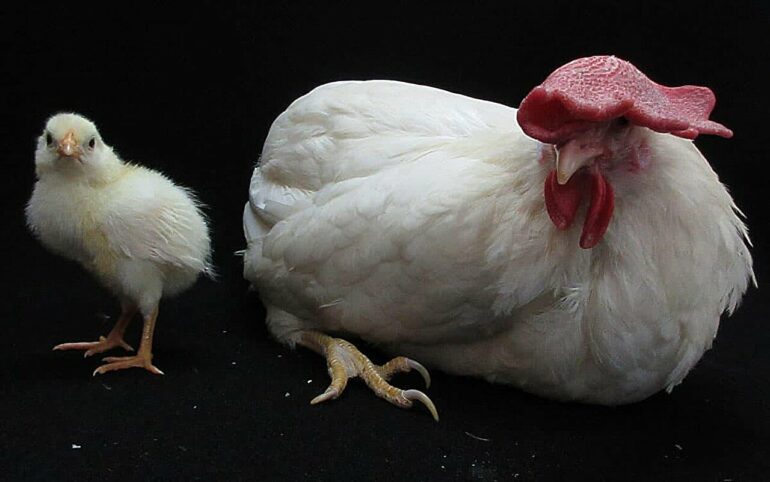
Furthermore, as these embryos continued to be cultured using the conventional shell-less method, embryonic development progressed, leading to the successful hatching of several normal chicks on the 21st day after the start of incubation.
The results of this research are expected to provide a foundation for various applications, including chick embryo development, toxicity assessments, and regenerative medicine.

More information:
Katsuya Obara et al, Real-time visualisation of developing chick embryos cultured in transparent plastic films from the blastoderm stage until hatching, Scientific Reports (2024). DOI: 10.1038/s41598-024-72004-y
Provided by
University of Tsukuba
Citation:
New method enables real-time visualization of chick embryo development from egg to chick (2024, October 15)

