Our cells have a complex relationship with the body’s microenvironment. It has been studied in the lab, but, to date, most studies leave out dynamic changes to the microenvironment. In other words, they look at cell life in a static world.
To better represent the reality of cell life in the ever-changing human body, researchers from TU/e have grown cells on hydrogels whose surface geometry can be changed on demand, at the micrometer scale, and repeatedly reshaped by illuminating it with blue light. Findings from the research could aid the development of future biomaterials to heal the body. The new research is published in the journal Advanced Science.
Life as a cell can be an arduous one. When it’s not worrying about invaders such as viruses or nasty bacteria, it has to contend with the ever-changing microenvironment, especially changes in the geometry of the surfaces upon which it resides.
“A cell’s microenvironment is a busy place. There’s the extracellular matrix (ECM)—the complex network made up of fibers, sugar, and water—and other cells of course,” says Maaike Bril, Ph.D. researcher in the Department of Biomedical Engineering. “Together, these influence the cell’s function and overall lifecycle.”
This constant interaction between cells and their microenvironment, which is better known as dynamic reciprocity, is complex, to say the least. “Through the interactions, cells and the environment are continually adapting their structures, geometries, and biophysical properties,” says Nicholas Kurniawan, assistant professor in the Department of Biomedical Engineering. “Yet, there’s a lot that remains to be learnt about how cells respond to geometric changes in the microenvironment”
Shine a blue light
There have been plenty of laboratory studies on how cells respond to changes in their microenvironment. However, many studies are missing one key aspect.
“Previous studies tend to focus on static settings where cells are placed on substrates that can be geometrically changed but remain in that configuration for the duration of the experiment,” says Bril. “This is not representative of the real microenvironment where the tissue shape changes continually.”
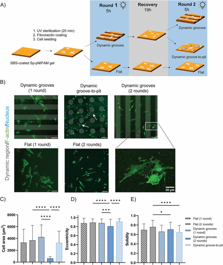
To capture this aspect, Bril, Kurniawan, and other TU/e colleagues have developed a cell-culturing substrate whose shape or surface topography can be changed using light, which has been published in Advanced Science.
“The substrate is made from a photoresponsive spiropyran-based hydrogel. When illuminated with blue light (wavelength of 455 nanometers) with specific spatial patterns, the substrate changes shape as the hydrogel swells, following those guiding patterns, and changes to the hydrogel are almost immediate,” says Bril. “In addition, the changes are reversible, meaning the hydrogel can used to study the effect of various surface topographies on cells.”
Redesigning hydrogels
Before the researchers could grow cells on the light-responsive hydrogels, the researchers had to solve one significant problem. “We had to re-design the hydrogel before we could grow cells on it,” says Bril.
To assist in the design of the hydrogel, Bril and Kurniawan reached out to the group of Albert Schenning (professor at the Department of Chemical Engineering and Chemistry) who specialize in the development of materials whose properties can be changed in response to light.
“Schenning’s team performed their previous research in water and at room temperature. However, for cells to grow, the conditions are different; they need to grow on a surface, in a medium that promotes growth, they need oxygen, and they need a humidified atmosphere at a temperature of about 37°C—the core temperature of the human body,” says Kurniawan.
However, it turned out that the chemical composition of the culture medium affected the hydrogel’s response to light, which explains why the researchers had to develop a new hydrogel specifically to create an environment to help cells grow. “To solve this, we included a layer of elastic rubber that shields the hydrogel from the chemicals in the cell culture medium that affected the way the hydrogel responds to light,” says Bril.
Fibroblasts remember
In terms of the type of cells they wanted to study with their new method, the researchers focused on fibroblast cells. These are cells that are responsible for the maintenance of skin, different tissues, and our organs. And, they made an exciting discovery.
“We discovered that the fibroblasts can ‘remember’ the past shape changes that have happened to the surfaces upon which they resided, and this is a completely new insight,” says Bril excitedly. “This memory of the past dynamics allows the cell to change how it functions to suit its needs. This could be a hugely important thing to consider when designing future biomaterials or better treatments to assist wound healing.”
Now that the researchers know how cells respond to changes in surfaces, they’d next like to learn more about what’s going on inside cells as they respond to shape changes in their environment.
“It would be great to know which proteins made inside a cell are essential for shape changes. And to study how they influence a so-called mechanical memory, or in this case topographical memory,” says Bril.
Added to that, the researchers plan to study how cells interact with other cells in greater detail because, in reality, cells in the body are not alone. “Integral to the life of a cell is interaction and communication with neighboring cells,” adds Bril.
Beyond probing cell-cell interactions further, the researchers also want to see how their research could be applied in new and exciting ways. “Shape-morphing hydrogels that support a living cell-matrix could have lots of applications that are waiting to be explored. For example, we could use them to mimic how tissue changes shape and size during growth,” says Kurniawan.
More information:
Maaike Bril et al, Shape‐Morphing Photoresponsive Hydrogels Reveal Dynamic Topographical Conditioning of Fibroblasts, Advanced Science (2023). DOI: 10.1002/advs.202303136
Provided by
Eindhoven University of Technology
Citation:
Redesigning hydrogels to shine a new light on cell responses in the body (2023, September 29)


