When Courtney “CJ” Johnson pulls up footage from her Ph.D. dissertation, it’s like she’s watching an attempted break-in on a home security camera.
The intruder cases its target without setting a foot inside, looking for a point of entry. But this intruder is not your typical burglar. It’s a virus.
Filmed over two and a half minutes by pinpointing its location 1,000 times a second, the footage shows a tiny virus particle, thousands of times smaller than a grain of sand, as it lurches and bobs among tightly packed human intestinal cells.
For a fleeting moment, the virus makes contact with a cell and skims along its surface but doesn’t stick before bounding off again. If this were an actual home break-in, Johnson says, “this would be the part where the burglar has not broken the window yet.”
Johnson is part of a Duke University team led by assistant chemistry professor Kevin Welsher. Together with Welsher’s postdoctoral associate Jack Exell and colleagues, they have come up with a way to capture real-time 3D footage of viruses as they approach their cellular targets. Their research is published today in the journal Nature Methods.
We inhale, ingest and take in millions of viruses every day. Most of them are harmless, but some of them—such as the viruses that cause the flu or COVID-19—can make us sick.
Infection starts when a virus binds to and enters a cell, where it hijacks the cellular machinery to make copies of itself. But before it can break in, a virus has to reach the cell first, Johnson said.
That often means getting through the protective layer of cells and mucus that line the airways and the gut—one of the body’s first lines of defense against infection.
The researchers wanted to understand how viruses breach these frontline defenses. “How do viruses navigate these complex barriers?” Welsher said. But these critical early moments before infection begins have long been difficult if not impossible to watch with existing microscopy methods, he added.
Part of the reason is that viruses move two to three orders of magnitude faster in the unconfined space outside the cell, compared with its crowded interior. To make things even trickier from an imaging perspective, viruses are hundreds of times smaller than the cells they infect.
“That’s why this is such a hard problem to study,” Johnson said. Under the microscope, “it’s like you’re trying to take a picture of a person standing in front of a skyscraper. You can’t get the whole skyscraper and see the details of the person in front of it with one picture.”
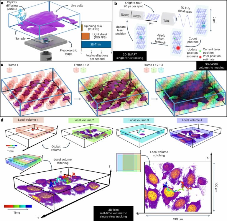
So the team developed a new method called 3D Tracking and Imaging Microscopy (3D-TrIm), which essentially combines two microscopes in one. The first microscope “locks on” to the fast-moving virus, sweeping a laser around the virus tens of thousands of times per second to calculate and update its position. As the virus bounces and tumbles around in the soupy exterior of the cell, the microscope stage continuously adjusts to keep it in focus.
While the first microscope tracks the virus, the second microscope takes 3D images of the surrounding cells. The combined effect, Welsher said, is akin to navigating with Google Maps: it not only shows your current location as you drive, it also shows the terrain, landmarks and the overall lay of the land, but in 3D.
“Sometimes when I present this work people ask, ‘is this a video game or a simulation?'” said Johnson, now a postdoctoral associate at the Howard Hughes Medical Institute Janelia Research Campus. “No, this is something that came from a real microscope.”
With their method the researchers can’t just, say, watch a healthy person breathe in virus particles from an infected person’s cough or sneeze. For one, they have to attach a special fluorescent label to a virus before they can track it—what the microscope follows is the movement of the glowing spot. And currently they can only track a virus for a few minutes at a time before it goes dim.
“The biggest challenge for us now is to produce brighter viruses,” Exell said.
But Welsher said he hopes the technique will make it possible to follow viruses in action beyond the coverslip, and in more realistic tissue-like environments where infections first take hold.
“This is the real promise of this method,” Welsher said. “We think that’s something we have the possibility to do now.”
More information:
Courtney Johnson et al, Capturing the start point of the virus–cell interaction with high-speed 3D single-virus tracking, Nature Methods (2022). DOI: 10.1038/s41592-022-01672-3
Citation:
Watch a virus in the moments right before it attacks (2022, November 11)


