
The human brain contains approximately 87 billion neurons. On average, each of these cells make thousands of different connections to facilitate communication across the brain. Neural communication is thought to underlie all brain functions – from experiencing and interpreting the world around you to remembering those experiences and controlling how your body responds.
But in this vast network of neural communication, precisely who is talking to whom, and what is the consequence of those individual conversations?
Understanding the details surrounding neural communication and how it’s shaped by experience is one of the many focuses of neuroscience. However, this is complicated by the sheer number of microscopic connections there are to study in the human brain, many of which are often in flux, and that available tools are unable to provide adequate resolution.
As a consequence, many scientists like me have turned to simpler organisms, such as the fruit fly.
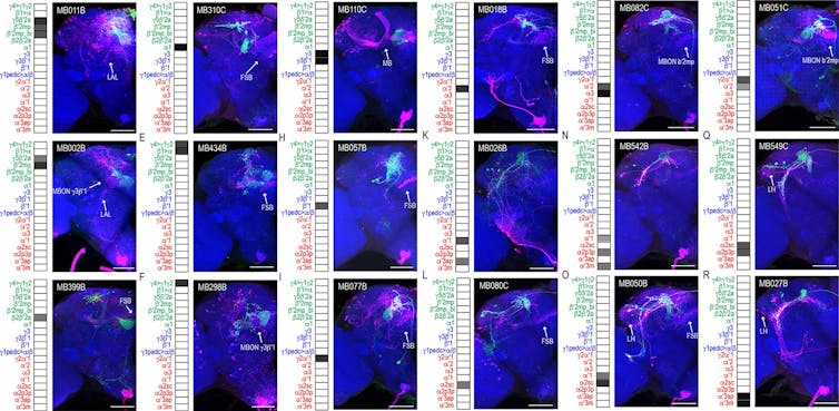
This figure shows connections between different mushroom body neurons.
Scaplen et al. 2021/eLife, CC BY
Fruit flies, though pesky in the kitchen, are invaluable in the laboratory. Their brains are built in remarkably similar ways to those of humans. Importantly, scientists have developed tools that make fly brains significantly easier to study with a resolution that hasn’t been achieved in other organisms.
My colleague Gilad Barnea, a neuroscientist at Brown University, and his team spent over 20 years developing a tool to visualize all of the microscopic connections between neurons within the brain.
Neurons communicate with each other by sending and receiving molecules called neurotransmitters between receptor proteins on their surface. Barnea’s tool, trans-Tango, translates the activation of specific receptor proteins into gene expression that ultimately allow for visualization.

My team and I used trans-Tango to visualize all the neural connections of a learning and memory center, called the mushroom body, in the fruit fly brain.
The video starts close to the face of the fly and moves back, using genetics to express different proteins within neurons to visualize them. Green indicates the neuron of interest, red indicates the neuron it talks to and blue indicates all other brain cells.
Kristin Scaplen, CC BY-SA
Here, a cluster of approximately four neurons, labeled green, receive messages from the mushroom body, which is the L-shaped structure labeled blue in the center of the fly brain. You can step through the brain and see all the other neurons they likely communicate with, labeled red. The cell bodies of the neurons reside on the edges of the brain, and the locations where they receive messages from the mushroom body appear as green tangles invading a small oval compartment. Where these weblike green extensions mingle with red are thought to be where neurons communicate their…

