UNSW scientists have grown human pancreatic cancer tumors in the lab—their model is the first of its kind, with important future clinical implications.
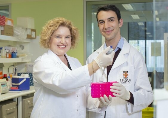
A team of medical researchers at UNSW have achieved a unique milestone in the fight against pancreatic cancer: led by world-leading cancer biologist Associate Professor Phoebe Phillips, the multi-disciplinary team has successfully grown a complete human tumor model in a petri dish.
Crucially, the team’s model stays intact for 12 days and offers a complete view of the tumor—an approach that has great potential for testing the effect of different drugs on the cancer, and offering personalized medicine approaches to patients down the track.
The findings are published today in Scientific Reports.
UNSW Scientia Ph.D. student John Kokkinos, who was set the challenge to create such a model by A/Prof. Phillips at the outset of his Ph.D., says current tumor models are limited. John was grateful to receive a Tour de Cure Ph.D. support scholarship to complete this project.
“One of the current gold standard models for testing therapeutics is the mouse—you give them pancreatic cancer and then test different treatments, but mouse tumors do not perfectly mimic the biology of the disease in patients,” Kokkinos says.
“Our ambitious vision—and the project I’ve been focused on for three years—was to take a human pancreatic tumor and keep it alive in a dish. If we could do that, we could use it to test which chemotherapeutics a patient’s tumor may respond best to.”
Replicating the cancer ‘fortress’
The unique advantage of the team’s approach is the fact that they didn’t just grow the tumor cells, but the tumor’s surrounding area, too. That makes it very different from the usual scientific approach of using so-called organoids.
“One of the hottest topics in cancer research is organoids, which basically involves taking tumor cells from a patient, isolating them, and then allowing them to form little 3-D tumor masses,” Kokkinos says.
“And so you’re left with just the tumor cells, and you’ve got no other surrounding cells, such as immune cells, scar tissue, blood vessels—which are all really critical in promoting the aggressiveness of the tumor.
“Think of these surrounding cells as the fortress that promotes tumor growth—it can really be a key player in driving resistance to chemotherapy.
“That’s why we felt that looking at tumor cells by themselves doesn’t really represent the true clinical picture.”
To keep everything intact, the team took the surgical piece of pancreatic tumor and developed a way to keep it alive for 12 days in an incubator in the laboratory.
“So essentially, we are trying to mimic the tumor in a way that best allows us to test therapeutics,” A/Prof. Phillips says.
“This is the first model of its kind that lasts this long—other labs have done something similar, but only for two or three days, and even then it doesn’t quite maintain the viability and the architecture of the tumors.
“We also characterized the tumor’s different cell types over time, and were able to show that these cell types don’t change. They maintain all of their characteristics in that 12-day period.”
While the team would love to be able to keep the tumor alive even longer, 12 days seems to be the current maximum.
“When we get the tumors from the patient, they are grown on a scaffold—that scaffold starts to degrade, and so we’re stuck with that time point,” A/Prof. Phillips says.
“But we want to work with our engineering colleagues to modify that scaffold to increase its life.”
Potential for personalized medicine
The end goal of developing such a model is to be able to test the effect of different drugs—both broadly and in terms of how they work on individual patients’ tumors.
“What it will allow us to do is test up to 10 different drugs simultaneously on a surgical specimen,” Kokkinos says.
“Because you get the result in a couple of weeks, you could go back and inform the clinical team about which drug is working best on a particular patient’s tumor—we hope that it’ll end up being a really rapid way to feed back into the clinical situation.”
Milestone on a long journey
Reflecting on the journey that got the team to this milestone, Kokkinos says it’s been a process of trial and error.
“It was a bit of a risk to start off with, I must say—we didn’t really know how it was going to turn out. There were a lot of failures along the way, things weren’t working out, but we kept persevering: we tried different things, we constantly went back to the drawing board.”
He remembers the first time he realized their idea might just work. “I must say the first time we actually got it right and we saw that the tumor architecture and the viability was maintained for 12 days, that was something quite extraordinary.
“And then from there, it was about characterizing the model, and looking at the individual cells that make up these mini tumors that we were growing, and then starting to test both the clinical drugs and the novel drugs, including our nanomedicine.”
Next step: more research, more patients
In today’s publication, the team describe in detail how their model works, and they’ve tested reproducibility across a couple of dozen patients. Now, they need funding to gather more data before this could be used as a clinical tool.
“Now, we want to actually show that our model does predict patient response accurately. That’s the next stage for us,” A/Prof. Phillips says.
The team also want to address one of their model’s biggest limitations: so far, they’ve exclusively worked on tumors from patients that had surgery.
“That represents about 15 to 20 percent of patients with pancreatic cancer, as the vast majority of patients don’t have a tumor that’s surgically resectable. So what we really want to try and do is be able to obtain tumor samples from patients that have metastatic disease, so that we can include all of the patients that have pancreatic cancer,” Mr Kokkinos says.
“So we’re looking into ways to do that—we are collaborating with clinicians and gastroenterologists to be able to take samples from a biopsy, and hopefully be able to use the model for those patients as well.”
Potential for national impact and global collaboration
The team say their model has the potential to be used globally.
“We think that this is a relatively simple model that can be adapted by multiple labs around the world,” Kokkinos says.
“They can take this model and use it on developing their own drugs. We’re also hoping this will enable us to establish new collaborations with other leading researchers.”
Pancreatic cancer has a five-year survival rate of only 10.7 percent, and is expected to become Australia’s second leading cause of cancer mortality by 2025—which is why it’s high on the agenda of health authorities in Australia, too: for example, A/Prof. Phillips is part of an advisory committee working on Australia’s National Pancreatic Cancer Roadmap to support improved outcomes and survival for people with pancreatic cancer, which Cancer Australia and the Department of Health are leading. She is also the co-lead of UNSW’s Pancreatic Cancer Research Hub, Australia’s first-ever center dedicated to preventing, treating, and eventually curing a cancer.
Disrupting the cellular process that promotes pancreatic cancer’s deadly growth
More information:
John Kokkinos et al. Ex vivo culture of intact human patient derived pancreatic tumor tissue, Scientific Reports (2021). DOI: 10.1038/s41598-021-81299-0
Provided by
University of New South Wales
Citation:
Human pancreatic cancer model offers new opportunities for testing drugs (2021, January 22)
retrieved 24 January 2021
from https://medicalxpress.com/news/2021-01-human-pancreatic-cancer-opportunities-drugs.html
This document is subject to copyright. Apart from any fair dealing for the purpose of private study or research, no
part may be reproduced without the written permission. The content is provided for information purposes only.


