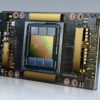
A miniature fluorescence microscope that weighs less while offering high resolution compared to existing devices will have a range of applications in systems biology. Existing miniature fluorescence microscopes are a standard technique in life sciences, but they only offer two-dimensional (2-D) information. In a new report now on Nature Light: Science & Applications, Kyrollos Yanny, Nick Antipa and a team of scientists in the Joint Graduate Program in Bioengineering, Electrical Engineering and Computer Sciences at the University of California, Berkeley and the Universite libre de Bruxelles Belgium, developed a single-shot 3-D fluorescence microscope. They engineered the new device known as the Miniscope3D by replacing the tube lens of a conventional 2-D miniscope with an optimized multifocal phase mask at the objective’s aperture stop. Using the device, Yanny and Antipa et al. optically recorded neural activity in free-moving animals and in long-term in situ imaging applications in incubators and within lab-on-a-chip devices.
Miniature fluorescence imaging and technical innovations
Miniature fluorescence microscopes are important in systems biology for optical recordings of neural activity in free-moving animals, long-term in situ imaging in incubators and medical devices. Such microscopes are also known as “miniscopes” and are made of 3-D printed parts, although offering 2-D fluorescence imaging alone. Single-shot methods can enable faster capture speeds and a temporal resolution limited by the camera frame rate. For example, a previously developed miniature light-field microscope (MiniLFM) can process neural activity with an optimized algorithm. In this work, Yanny et al. developed a 3-D miniscope to achieve higher resolution with lighter weight compared to existing techniques. The team tested the microscopic capabilities by imaging fluorescent resolution targets as well as freely swimming biological samples and mouse brain tissue. They validated the reconstructed outcomes in comparison with two-photon microscopy to understand limits of the new technique.
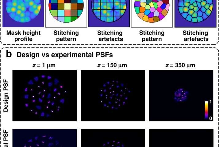
To achieve high-quality imaging in a small, low-weight device, Yanny et al. placed the phase mask (where light passing through the mask will undergo a phase-shift proportional to the thickness of the material) in Fourier space to reduce computational burden and improve compactness. They added 3-D capabilities to the 2-D miniscope at the cost of a small loss in lateral resolution and lower signal-to-noise ratio. The algorithm united the optical theory with compressed sensing to fabricate the optimized phase masks. The technique facilitated a new miniature 3-D microscope architecture with higher resolution, open-source designs, higher-quality fabrication and an efficient calibration scheme or reconstruction algorithm.
Characterizing the computational microscope and investigating the mouse brain
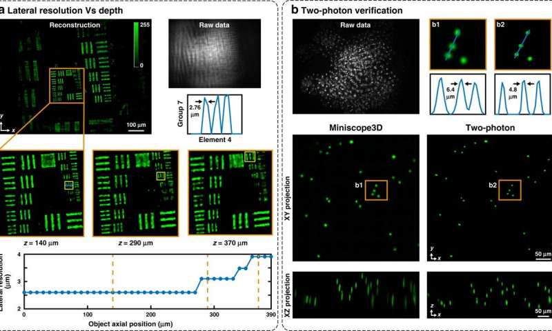
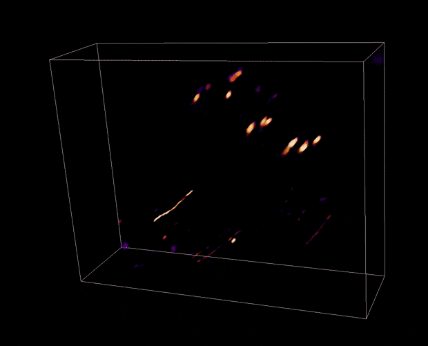
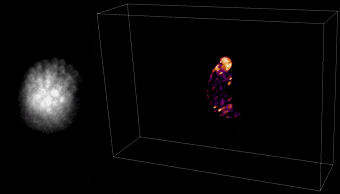
The team tested the performance of the computational microscope using samples of increasing complexity to capture 3-D dynamic recordings. They measured the lateral resolution at different depths by imaging a fluorescent resolution target. They then validated the accuracy of their results using two-photon microscopy. For example, the Miniscope3D could accurately recover all reconstructed images of the 3-D fluorescent bead sample post-processing. They showed the potential of the method using neuro-biological samples where green fluorescent protein tagged regions expressed sparse populations of neurons throughout the sample. The reconstructed images obtained from different parts of the hippocampus showed dendrites running across the surface alongside individual cell bodies. When Yanny et al. next investigated dynamic samples of free-swimming, green-dyed tardigrades (also known as water bears), the reconstructed images showed the efficiency of Miniscope3D imaging to track freely moving biological organisms at high resolution in space-time.
Applications and accessibility of the device
Most applications of Miniscope3D will be similar to 3-D microscopy and MiniLFM (miniature light-field microscopy), which is considered the gold standard for single-shot miniature 3-D fluorescence imaging. Compared to MiniLFM, however, the new Miniscope3D method offered multiple improvements including multifocal lenses, best—case lateral resolution and a 10-fold increase in the useable measurement volume. The improved performance arrived in a hardware package smaller than the MiniLFM with lighter weight to freely observe moving organisms. The method further enabled experimental reconstruction with or without scattering for mouse brain tissue at single neuron resolution. The team will optimize existing limits of the device including scattering, for further applications.
By building upon a popular open-source miniscope platform, Yanny et al. provided accessibility for the Miniscope3D design. In this way, Kyrollos Yanny, Nick Antipa and colleagues provided a 3-D prototype as an opportunity to upgrade the 2-D miniscopes currently in use across 450 laboratories. The experimental results were in good agreement with the theoretical design and analysis to serve as a useful framework for customized single-shot 3-D systems.
High-resolution and large field-of-view Fourier ptychographic microscopy
More information:
Kyrollos Yanny et al. Miniscope3D: optimized single-shot miniature 3-D fluorescence microscopy, Light: Science & Applications (2020). DOI: 10.1038/s41377-020-00403-7
Kunal K Ghosh et al. Miniaturized integration of a fluorescence microscope, Nature Methods (2011). DOI: 10.1038/nmeth.1694
Jesse K. Adams et al. Single-frame 3-D fluorescence microscopy with ultraminiature lensless FlatScope, Science Advances (2017). DOI: 10.1126/sciadv.1701548
2020 Science X Network
Citation:
Miniscope3D—A single-shot miniature three-dimensional fluorescence microscope (2020, October 15)
retrieved 15 October 2020
from https://phys.org/news/2020-10-miniscope3da-single-shot-miniature-three-dimensional-fluorescence.html
This document is subject to copyright. Apart from any fair dealing for the purpose of private study or research, no
part may be reproduced without the written permission. The content is provided for information purposes only.

