A joint research team from DGIST led by Professors Jin Ho Chang and Jae Youn Hwang of the Department of Electrical Engineering and Computer Science has developed the world’s first laser scanning microscopy technology that enables deeper and more detailed observation of biological tissues using gas bubbles that are temporarily produced by ultrasound.
Optical imaging and therapeutic technologies are widely utilized in life science research and clinical practice. However, due to the occurrence of optical scattering in the tissue, the light transmission is low. Consequently, there exists an inherent limitation in the image acquisition and treatment of deep tissue. This significantly hinders the expansion of the application field.
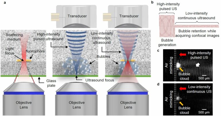
To overcome this, in 2017, Professor Jin Ho Chang’s team envisioned that it would be possible to use micrometer-sized gas bubbles that are normally observed when tissues are exposed to high-intensity ultrasound. They developed a technology based on the fact that gas bubbles temporarily created by ultrasonic waves cause optical scattering in the same direction as the propagation of incident light, thus increasing the penetration depth of light.
Furthermore, the joint research team of Professors Jin Ho Chang and Jae Youn Hwang focused on expanding the application of optical imaging technology using ultrasound-induced gas bubbles. A confocal fluorescence microscope is a device that selectively detects fluorescence signals generated at the focal plane of light and provides high-resolution, high-contrast images of microstructures such as cancer cells. It is the most widely used device in life science research owing to its high performance. However, the focus of the light is blurred at depths exceeding 100 µm owing to the scattering of light that occurs inside the tissue, which significantly limits the application and effectiveness of confocal fluorescence microscopy.
To increase the maximum imaging depth of optical imaging modalities, such as confocal fluorescence microscopy, photons constituting the irradiated light must not have a phenomenon in which their propagation direction is distorted by light scattering in tissues. However, the previously developed method based on gas bubbles sparsely created by ultrasound was not a solution.
Therefore, this joint research team developed ultrasound technology to create a bubble layer in the desired area with dense gas bubbles (with a density of 90% or more) inside living tissue and to maintain the generated gas bubbles while acquiring the image. In this gas bubble layer, the distortion in the propagation direction of photons does not occur.
Therefore, it has been experimentally proven that light focusing is possible even in deeper biological tissues. In addition, by applying this technology (i.e., ultrasound-induced tissue transparency) to a confocal fluorescence microscope, the UltraSound-induced Optical Clearing Microscopy (US-OCM), named in this study, was developed for the first time in the world with an imaging depth six times longer than that of the conventional confocal microscopy.
In particular, the US-OCM developed in this study did not cause any damage to the tissue because when the ultrasonic irradiation was stopped, the generated gas bubbles disappeared, and the optical properties returned to before gas bubble generation, suggesting that it is harmless to the living body.
Professor Jin Ho Chang of the Department of Electrical Engineering and Computer Science at DGIST says that “through close collaboration with ultrasound and optical imaging experts, we were able to overcome the inherent limitations of existing optical imaging and treatment technologies.”
“The technology secured through this study will be applied to various optical imaging techniques including multiphoton microscopy and photoacoustic microscopy in addition to several optical therapies including photothermal therapy and photodynamic therapy. This would enhance the application of existing technologies by increasing their image and treatment depth.”
The results of this research were published in Nature Photonics.
More information:
Haemin Kim et al, Deep laser microscopy using optical clearing by ultrasound-induced gas bubbles, Nature Photonics (2022). DOI: 10.1038/s41566-022-01068-x
Provided by
DGIST (Daegu Gyeongbuk Institute of Science and Technology)
Citation:
Medical optical imaging using the world’s first ‘ultrasound-induced tissue transparency’ technology (2022, October 7)


