Viral infections have challenged humanity for centuries. Even with progressive scientific advancements, the struggle against viruses continues, as exemplified by the recent COVID-19 pandemic. In the fight against these viral infections, a variety of techniques have been established for detecting and quantifying viruses, contributing significantly to the development of critical vaccines and antiviral medications.
Among these techniques, the viral plaque assay stands out as the gold standard due to its unique ability to assess virus infectivity in a cost-effective way by observing the formation of viral plaques caused by viral infections over a layer of cells. Nevertheless, the traditional viral plaque assays require an incubation period of 2–14 days, followed by sample staining using chemicals and human visual inspection to count the number of viral plaques.
This procedure is time-consuming and susceptible to staining artifacts and counting errors induced by human technicians. Therefore, an accurate, automated, rapid, and cost-effective viral plaque quantification technique is urgently needed.
In a new paper published in Nature Biomedical Engineering, a team of scientists led by Professor Aydogan Ozcan from the Electrical and Computer Engineering Department at the University of California, Los Angeles (UCLA), developed a rapid, stain-free and automated viral plaque detection system enabled by holography and deep learning.
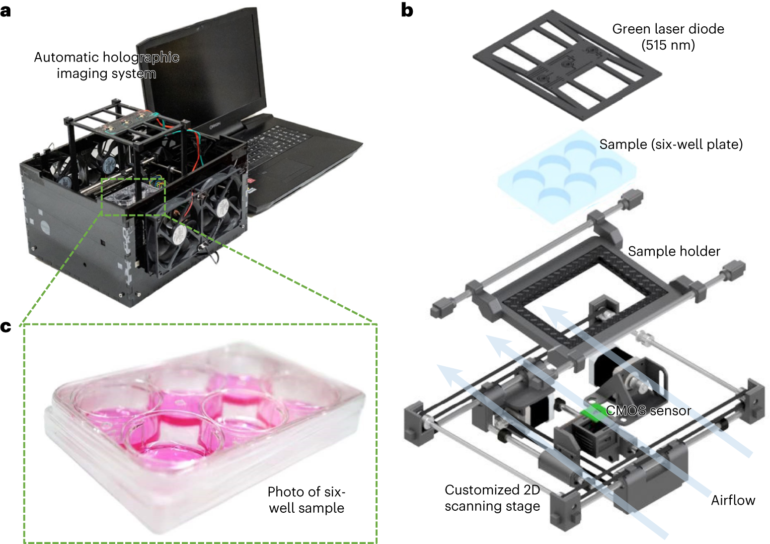
This system incorporates a cost-effective and high-throughput holographic imaging device that continuously monitors the unstained virus-infected cells during their incubation process. At each imaging cycle, these time-lapse holograms captured by the device are periodically analyzed by an AI-powered algorithm to automatically detect and count the viral plaques that appear due to virus replication.
The proof-of-concept and effectiveness of this system were demonstrated using three different types of viruses: vesicular stomatitis virus (VSV), herpes simplex virus type-1 (HSV-1) and encephalomyocarditis virus (EMCV). By utilizing this system, UCLA researchers achieved the detection of more than 90% of VSV viral plaques within 20 hours of incubation without any chemical staining, demonstrating a time saving of more than 24 hours in comparison to the traditional plaque assay, which requires 48 hours of sample incubation. In the case of HSV-1 and EMCV, this system effectively reduced their viral plaque detection times by approximately 48 and 20 hours, respectively, compared to the detection time needed for the traditional staining-based viral plaque assay.
In addition to offering major time savings, this stain-free and cost-effective system can successfully identify individual viral plaques within clusters as opposed to the traditional viral plaque assays, which fail to separately detect and count those individual plaques within clusters due to the spatial overlap of their signatures.
All these findings highlight the transformative potential of this AI-powered viral plaque detection system to be used with various plaque assays in virology, which might help to expedite vaccine and drug development research by significantly reducing the detection time needed for traditional viral plaque assays and eliminating chemical staining and manual counting entirely.

More information:
Tairan Liu et al, Rapid and stain-free quantification of viral plaque via lens-free holography and deep learning, Nature Biomedical Engineering (2023). DOI: 10.1038/s41551-023-01057-7
Provided by
UCLA Engineering Institute for Technology Advancement
Citation:
Deep learning and holographic imaging accelerate the detection and quantification of viral plaques (2023, June 23)

