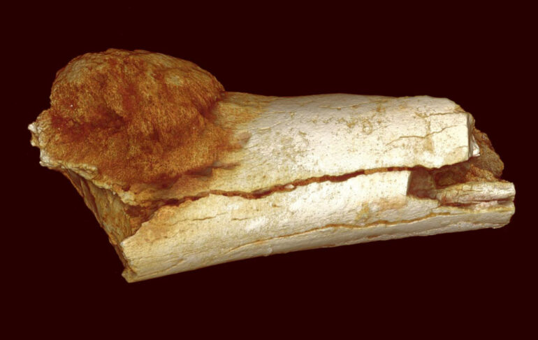
More than 215 million years ago, a large amphibian species lived in floodplains in southwestern Poland: Metoposaurus krasiejowensis. On one of these fossils, Polish and American scientists, with the participation of researchers from the University of Bonn, detected bone cancer for the first time. The results have now been published in the journal BMC Ecology and Evolution.
Traces of diseases occasionally found in the bones of prehistoric animals are evidence of the ancient ancestry of some diseases. Now, an interdisciplinary, international research team led by Dr. Dawid Surmik of the Faculty of Natural Sciences at Silesian University in Katowice, Poland, has produced another important piece of evidence for the occurrence of cancers in Earth’s distant past.
In the article, the scientists present the results of their study of a vertebra. It originates from the Upper Triassic and can be traced back to an extinct Temnospondyl amphibian: Metoposaurus krasiejowensis. The fossil was discovered in the famous Krasiejów locality located near Opole in southwestern Poland, known for numerous remains of fossil vertebrates.
Pathological tissue penetrated deep into the vertebra
The researchers noticed an overgrowing bone mass covering a large part of the vertebra, which belongs to the scientific collection of the Institute of Paleobiology of the Polish Academy of Sciences in Warsaw. Working with the University of Silesia’s Faculty Laboratory of Computational Microtomography, the researchers used X-rays to examine the internal structure of this fossil. The scans showed that the diseased tissue not only grew around the vertebra from the outside, but also penetrated deep into its interior.
“Based on this data, it became clear that the cause of the overgrowth was a malignant tumor,” Surmik says. Further analysis of the scans revealed that much of the vertebra’s original structure had been destroyed by the growth of cancerous pathological tissue. The researchers sectioned the sample and produced a thin, translucent sample that can be viewed in a transmitted light microscope—much like modern cancer samples are examined in medical laboratories.

Reconstruction of growth and stages of tumour invasion. © Jakub Zalewski
Histological analyses at the University of Bonn
Paleontologists Sudipta Kalita, Elzbieta M. Teschner and Dr. Dorota Konietzko-Meier of the Institute of Geosciences at the University of Bonn performed the histological analyses and three-dimensional reconstruction of the vertebra.
“These data helped us make a kind of ‘medical’ diagnosis,” Dr. Konietzko-Meier says. “We proved that the bone tissue took an atypical course and were able to describe the specific tumor development and reconstruct its ontogenetic growth dynamic, thus how the tumor grew during the animals’ life. This was the first time when the growth dynamic was described for an extinct animal.”
The dissected vertebral specimen revealed a lot of information about the histological structure of the pathological tissue, especially about the contact between the cancerous part and the healthy one. Based on these observations, the research team concluded that the cancer that had affected the fossil amphibian was osteosarcoma. It is among the most common malignant bone tumors in humans.
“This malignant tumor in Metoposaurus is currently one of the oldest examples of cancer in the fossil record and the only one associated with a fossil amphibian,” Surmik says. It is also the best-documented evidence of cancer in a prehistoric animal, he adds, supported by solid results from microstructural studies.
More information:
Dawid Surmik et al, An insight into cancer palaeobiology: does the Mesozoic neoplasm support tissue organization field theory of tumorigenesis?, BMC Ecology and Evolution (2022). DOI: 10.1186/s12862-022-02098-3
Provided by
University of Bonn
Citation:
A bone cancer tumor more than 215 million years old (2022, December 22)

