Of the three groups of rotavirus that cause gastroenteritis in people, called groups A, B and C, groups A and C affect mostly children and are the best characterized. On the other hand, of group B, which causes severe diarrhea predominantly in adults, little is known about the tip of the virus’s spike protein, called VP8* domain, which mediates the infection of cells in the gut.
“Determining the structure of VP8* in group B rotavirus is important because it will help us understand how the virus infects gastrointestinal cells and design strategies to prevent and treat this infection that causes severe diarrheal outbreaks,” said corresponding author Dr. B. V. Venkataram Prasad, professor of biochemistry and molecular biology at Baylor College of Medicine.
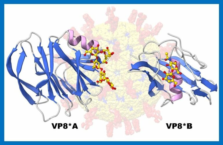
The team’s first step was to determine the 3D structure of VP8* B using X-Ray crystallography, a laborious and time-consuming process. However, this traditional approach was unsuccessful in this case. The researchers then turned to a recently developed artificial intelligence-based computational program called AlphaFold2.
“AlphaFold2 predicts the 3D structure of proteins according to their genetic sequence,” said first author and co-corresponding author Dr. Liya Hu, assistant professor of biochemistry and molecular biology at Baylor. “We knew that the protein sequence of VP8* of rotavirus group B was about 10% similar to the sequences of VP8* of rotavirus A and C, so we expected differences in the 3D structure as well. But we were surprised when AlphaFold2 predicted a 3D structure for the VP8* B that was not just totally different from that of the VP8* domain in rotavirus A and C, but also that no other protein before had been reported to have this structure.”
With this information in hand, the researchers went back to the lab bench and experimentally confirmed that the structure of VP8* B predicted by ALphaFold2 indeed coincided with the actual structure of the protein using X-ray crystallography.

Rotavirus particles visualized by immune electron microscopy. Notice the viral spike indicated by the arrow. © Centers for Disease Control.
How rotavirus infects cells
Previous research has shown that rotavirus A and C infect cells by using the VP8* domain to bind to specific sugar components on histo-blood group antigens, including the A, B, AB and O blood groups, present in many cells in the body. It has been proposed that the ability of different rotavirus to bind to different sugars on the histo-group antigens might explain why some of these viruses specifically infect young children while others affect other populations. Unlike the VP8* A and VP8* C, the sugar specificity of VP8* B had not been characterized until now.
“We screened VP8* B against an array of sugars and found that it recognizes N-acetyllactosamine, a sugar common in many cells in the body, that is not recognized by VP8* of rotavirus A and C,” Hu said. “Such a 3D structure that also is capable of binding to sugar has not been described before.”
“I am excited about identifying a novel 3D protein structure. I am also anticipating all the discoveries that will come from this as we investigate how the new structure interacts with cells to infect them and how this process compares to the one from rotavirus A and C,” said co-author Dr. Wilhelm Salmen, a postdoctoral fellow in the Prasad lab.
“Our lab has been collaborating with Dr. Prasad’s lab for many years to understand the significance of sugar-binding viruses in gastrointestinal infections,” said co-author Dr. Mary Estes, Cullen Foundation Endowed Chair and Distinguished Service Professor of Virology and Microbiology at Baylor. Estes also is a member of Baylor’s Dan L Duncan Comprehensive Cancer Center. “We can’t cultivate the group B virus yet, but our lab will now try to grow these adult viruses in our human organoid systems, a miniature model of the human gut that can help us probe the mechanism of virus entry and growth. This may lead to new therapeutics that are still needed to treat diarrheal disease.”
“This new approach to determine a protein’s 3D structure represents a significant step forward for the field of structural biology,” Hu said.

“I am excited about our findings of a new 3D protein structure from an evolutionary point of view. It exemplifies how viruses can evolve by incorporating structurally distinct modules with similar functionality, but how this structure came about in this group B rotavirus is quite intriguing,” said Prasad, who holds the Alvin Romansky Chair in Biochemistry and is a member of the Dan L Duncan Comprehensive Cancer Center.
The research was published in Communications Biology.
More information:
Liya Hu et al, Novel fold of rotavirus glycan-binding domain predicted by AlphaFold2 and determined by X-ray crystallography, Communications Biology (2022). DOI: 10.1038/s42003-022-03357-1
Provided by
Baylor College of Medicine
Citation:
Artificial intelligence reveals a never-before described 3D structure in rotavirus spike protein (2022, June 9)

