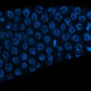
Visualizing cells after editing specific genes can help scientists learn new details about the function of those genes. But using microscopy to do this at scale can be challenging, particularly when studying thousands of genes at a time.
Now, researchers at the Broad Institute of MIT and Harvard, along with collaborators at Calico Life Sciences, have developed an approach that brings the power of microscopy imaging to genome-scale CRISPR screens in a scalable way.
PERISCOPE—which stands for perturbation effect readout in situ via single-cell optical phenotyping—combines two technologies developed by Broad scientists: Cell Painting, which can capture images and key measures of subcellular compartments at scale, and Optical Pooled Screening, which “barcodes” cells and uses CRISPR to systematically turn off individual genes to study their function in those cells.
The new technique lets scientists study the effects of perturbing over 20,000 genes on hundreds of image-based cellular features. Generating data with the method is more than 10 times less expensive than comparable high-dimensional approaches such as high-throughput single-cell RNA sequencing and can be adapted to study a wide variety of cell types.
In their article published in Nature Methods, the researchers applied PERISCOPE to execute three whole-genome CRISPR screens to create an open-source atlas of cell morphology.
The study is the result of a collaboration between the labs of Broad co-senior authors JT Neal, institute scientist and co-director of type 2 diabetes systems genomics; Anne Carpenter, institute scientist and senior director of the Imaging Platform; and Paul Blainey, core institute member; as well as Calico, led by co-senior author Calvin Jan. Broad staff scientists Meraj Ramezani and Erin Weisbart, research associate Julia Bauman, and postdoctoral researcher Avtar Singh were all co-first authors on the study.
“This atlas itself is a first-in-class genome-scale resource for linking cell morphology to gene function, and I think it has a lot of discovery potential,” said Neal. “But maybe more importantly, all of the data is open access, and we’ve built really robust industrial-grade analysis pipelines that can be repurposed by anybody to analyze optical pooled screening data at scale.”
Morphological insights
First developed by Carpenter in 2013, Cell Painting is an assay designed to image multiple cell organelles at the same time and at scale, “painting” them different colors such as nuclei blue and mitochondria red. Machine-learning models then detect hundreds of subtle changes in the images, such as the texture of the cytoplasm or area of the nucleus, allowing researchers to test the effects of a gene perturbation, for example, on the cell.
When Neal joined the Broad in 2017 as a researcher in the Cancer Program, he was intrigued by the possibility of applying Cell Painting to cancer cells to learn the function of key genes and variants, which could help in the development of targeted therapies. He struck up a conversation with Carpenter, and also with Blainey, whose lab had developed optical pooled screening.
The three started discussing how they might combine the two techniques and, in 2019, started building PERISCOPE, including an associated computational pipeline that integrates with Cell Profiler, in collaboration with Calico.
In PERISCOPE, the researchers introduce a library of guide RNAs targeting about 20,000 genes into cells. Next, they induce the expression of the DNA-cutting Cas9 enzyme to disable the genes targeted by the guides.
They then convert the guide RNAs to complementary DNA, creating “barcodes” of the gene knocked out in each cell, which allow scientists to identify which gene has been turned off in each cell. This barcoding also enables the study of many genes in a single batch of cells, which is more efficient than testing them one by one.
Discover the latest in science, tech, and space with over 100,000 subscribers who rely on Phys.org for daily insights.
Sign up for our free newsletter and get updates on breakthroughs,
innovations, and research that matter—daily or weekly.
Finally, they use a standard widefield microscope to record images of the five Cell Painting stains and the four-color barcodes in the cells, followed by automated image analysis to extract cell features and link them to guide RNAs.
Using PERISCOPE, the researchers created atlases of the effects of knocking out genes in human lung and cervical cancer cells in standard cell culture media as well as in media that more closely resembles the physiological environment.
These atlases not only illustrated known biology but also revealed new information about poorly characterized genes. For example, they uncovered the function of TMEM251, a gene that has been linked to a rare genetic lysosomal storage disease. The team found that it is required for trafficking enzymes to lysosomes.
Next, the team is working on building the capacity to image even more colors simultaneously, expanding the range of traits that PERISCOPE can capture. They’re also collaborating with researchers in Broad’s Novo Nordisk Foundation Center for Genomic Mechanisms of Disease to create perturbation atlases in cardiometabolic cell types, and with Broad’s Ladders to Cures Accelerator to identify treatments for rare genetic disorders.
In addition, the team anticipates that PERISCOPE will be applicable to neurodegenerative disorders, such as Parkinson’s disease. Neal says PERISCOPE could one day even enable studies of the interactions between multiple genes.
“In the past, that’s been tricky because the genetic interaction space scales very quickly,” he said. “With PERISCOPE and other scalable optical pooled screening approaches, it’s now no longer far-fetched to envision very large genetic interaction screens.”
More information:
Ramezani M et al, A genome-wide atlas of cell morphology, Nature Methods (2025). DOI: 10.1038/s41592-024-02537-7. www.nature.com/articles/s41592-024-02537-7
Provided by
Broad Institute of MIT and Harvard
Citation:
Genome-wide atlas of cell morphology reveals gene functions (2025, January 27)


