When it comes to oxygen, you can have too much of a good thing. Breathing air that contains higher levels of oxygen than the usual 21 percent found in Earth’s atmosphere can cause organ damage, seizures, and even death in people and animals, particularly if it’s in excess of the body’s oxygen needs. Until now, however, scientists have mostly speculated about the mechanisms behind this phenomenon, known as oxygen toxicity, or hyperoxia.
Now, researchers at Gladstone Institutes have discovered how excess oxygen changes a handful of proteins in our cells that contain iron and sulfur—a chemical process similar to the rusting of iron. In turn, those “rusty” proteins trigger a cascade of events that damage cells and tissues. The findings, published in the journal Molecular Cell, have implications for conditions such as heart attacks and sleep apnea.
“This study allowed us to put together a very specific timeline for what happens in hyperoxia,” says Gladstone Assistant Investigator Isha Jain, Ph.D., senior author of the new study. “The results weren’t at all what we were expecting, but it’s very interesting and exciting to now know how this sequence of events unfolds.”
An understudied question
At high levels, oxygen is toxic to every form of life, from bacteria and plants to animals and people. Of course, not enough oxygen is also fatal; there’s an intermediate, “Goldilocks” amount under which most life on Earth thrives—not too much and not too little.
While clinicians have long studied the details of how oxygen shortage impacts cells and tissues (for example, in heart attacks and strokes), the effects of excess oxygen have been relatively understudied.
“For many years, the medical teaching was that to a certain degree, more oxygen was better, or at least benign, when treating patients with conditions such as heart attacks,” says Alan Baik, MD, a postdoctoral scholar in Jain’s lab and a cardiologist at UC San Francisco (UCSF). “But there has now been a growing number of clinical studies showing that excess oxygen actually leads to worse outcomes. This motivated us to better understand why excess oxygen can be toxic.”
Studies have recently revealed, for instance, that breathing too much supplemental oxygen might be detrimental to heart attack patients and premature infants. Similarly, in obstructive sleep apnea, the sudden bursts of oxygen that follow pauses in breathing have been shown to be a key component of how the disorder increases patients’ risks of chronic health problems.
Still, the mechanisms of these effects remained murky. Many researchers assumed that reactive oxygen species—unstable and highly reactive oxygen derivatives that can damage our genome and many molecules in our cells—likely played a role in hyperoxia, but there was little evidence to demonstrate how excess oxygen affects specific enzymes and pathways.
How CRISPR found the answer
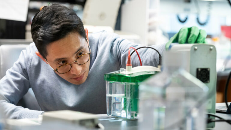
Jain’s group—including Baik, postdoctoral fellow Galih Haribowo, Ph.D., and graduate student Kirsten Xuewen Chen, who are co-first authors of the new paper—turned to the genome editing technology CRISPR to test the roles of a variety of genes in hyperoxia.
Using CRISPR, the researchers removed, one at a time, more than 20,000 different genes from human cells grown in the lab and then compared the growth of each group of cells at 21 percent oxygen and 50 percent oxygen.
“This kind of unbiased screen let us probe the contributions of thousands of different pathways in hyperoxia rather than just focusing on those we already suspected might be involved,” says Jain, who is also an assistant professor of biochemistry at UCSF. “It led us toward molecules that have never been uttered before in the same sentence as oxygen toxicity.”
Four molecular pathways stood out in the screen as being involved in the effects of hyperoxia. They related to diverse cellular functions including the repair of damaged DNA, the production of new DNA building blocks, and the generation of cellular energy.
Protein clusters in common
At first, the team couldn’t pinpoint what the four pathways had in common and why they were all impacted by high oxygen levels. It took some molecular sleuthing to discover that each pathway had a critical protein that contained iron atoms connected to sulfur atoms—so-called “iron-sulfur clusters”—in its molecular structure.

The researchers went on to show that in as little as 30 percent oxygen, the iron-sulfur clusters in the four proteins become oxidized—they chemically react with oxygen atoms—and that change causes the proteins to degrade. As a result, cells stop functioning correctly and consume even less oxygen, causing a further increase in oxygen levels in the surrounding tissues.
“One important takeaway is that hyperoxia is not impacting cells and tissues solely through reactive oxygen species, as many had assumed,” says Jain. “That means the use of antioxidants—which can combat reactive oxygen species to some degree—is unlikely to be sufficient to prevent oxygen toxicity.”
More information:
Alan H. Baik et al, Oxygen toxicity causes cyclic damage by destabilizing specific Fe-S cluster-containing protein complexes, Molecular Cell (2023). DOI: 10.1016/j.molcel.2023.02.013
Provided by
Gladstone Institutes
Citation:
Researchers discover how too much oxygen damages cells and tissues (2023, March 9)

