Exposure to mercury (Hg) is extremely neurotoxic in most chemical forms. Even scientists who study mercury compounds are at risk due to potential exposure to Hg. Renowned physicist Michael Faraday suffered from Hg poisoning due to prolonged exposure to Hg vapors, leading him to halt his research at the age of 49 due to deteriorating health. Another example is lab chemist Karen Wetterhahn who was killed by dimethylmercury poisoning after a few drops escaped from a pipette and landed on one of her latex-gloved hands.
Numerous studies have focused on the exposure and effects of Hg, particularly in marine and sea creatures. It is well-known that people should limit the consumption of certain fish, like tuna, due to mercury presence. However, the question arises: can mercury ions reach the brains of terrestrial animals?
Dr. Yulia Pushkar, a professor of Physics and Astronomy at Purdue University’s College of Science was initially skeptical. She has maintained a brain imaging program since 2008 at Purdue University. Her group, with expertise in sample preparation, measurements and data analysis, is sought after by researchers in U.S. and worldwide, including these from Japan and more recently Australia.
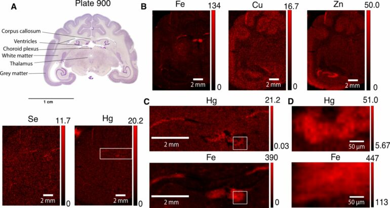
Pushkar’s research group was tasked with checking for Hg in brains of mongooses collected in Okinawa Island. Surprisingly, brain scans revealed mercury in these invasive animals. The research group refined the scans, achieving a resolution of a few tens of nanometers to observe the affected brain cells. Their collaborative findings were recently published in Environmental Chemistry Letters.
The mystery of how mercury enters the mongoose brain remains unresolved. Possible sources include the water they drink, bird eggs they consume, mineral exposure, or even the air they breathe. One thing is very clear though, this is a very bad sign.
“Hg is very toxic at low concentrations as Hg can bind and affect the function of essential biomolecules,” explains Pushkar. “Efficiency of detoxification will depend on uptake and binding constant inside detected accumulations and potential leakage from these if brain cells die. As of now, there is no known way to safely dissolve these aggregates from tissue and there are no reports of reversing Hg poisoning of neural system. The main approach we should all take is to avoid any exposures, especially chronic ones like in Faraday’s case.”
“I was skeptical whether any Hg could be detected. Usually, neurotoxic elements even if they get into brains are present in ultra-low concentrations,” explains Pushkar. “We took these specimens to the Advanced Photon Source at Argonne National Laboratory where brains were exposed to intense X-rays. Defying my skepticism, the Hg signal was present.”
Scanning across brain samples researchers started tracing brain areas which seemed to have higher Hg content. After three years of study and five trips to two national synchrotron facilities (Advanced Photon Source at Argonne National Laboratory and NSLS-II at Brookhaven National Laboratory) researchers can now report that particular brain cells: cells of choroid plexus (making up blood cerebrospinal fluid barrier) and astrocytes of the subventricular zone contain Hg rich puncta (~0.5–2 microns in size).
Pushkar’s team of researchers believes these cells help to filter Hg from the blood and brain tissue and store with the help of another element, Selenium (Se). Which particular Se containing biological molecules bind Hg remains to be discovered.
Pushkar’s team for this publication consists of Pavani Devabathini and Gabriel Bury (both graduate students) and then-undergraduate student Darrell Fischer (presently at Harvard graduate school). Data were collected by the entire team and analyzed by Devabathini and Fischer. Once the data were analyzed, the entire team contributed to the writing of the publication.
This discovery holds significance for environmental monitoring in terrestrial animals and provides new tools for tracing Hg in brain cells, potentially impacting human health and safety.
“Human activities result in the emission of 2,000 metric tons of mercury compounds annually and we do not fully understand where all this neurotoxic Hg ends up,” says Pushkar. “Most studies so far focused on marine biota (fish and whales) but apparently terrestrial species are also affected. We expect the human brain reacts to Hg in a similar fashion via interactions with cells of choroid plexus and astrocytes. However, we do not know if the human brain has enough Se-containing biomolecules to bind to Hg.”
More information:
Pavani Devabathini et al, High-resolution imaging of Hg/Se aggregates in the brain of small Indian mongoose, a wild terrestrial species: insights into intracellular Hg detoxification, Environmental Chemistry Letters (2023). DOI: 10.1007/s10311-023-01666-3
Citation:
High levels of mercury traced to particular cell types in brains of mammals (2024, January 4)


