Ultrafast imaging plays an important role in physics and chemistry to investigate the femtosecond dynamics of nonuniform samples. The method is based on understanding phenomena induced by an ultrashort laser pump pulse using an ultrashort probe pulse thereafter. The emergence of very successful ultrafast imaging techniques with an extremely high frame-rate is based on wavelength or spatial frequency encoding. In a new report now in Light: Science & Applications, Chen Xie, Remi Meyer, and a team of scientists in China and France used a pump-induced micrografting method to provide detailed in situ characterization of a weak probe pulse. The method is non-destructive and fast to perform and therefore the in-situ probe diagnostic can be repeated to calibrate experimental conditions. The technique will allow previously inaccessible imaging to become feasible across a field of superfast science at the micro- and nanoscale.
Superfast physics and chemistry
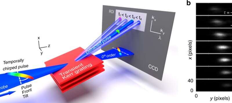
The concept of laser matter interactions in ultrafast physics and chemistry is based on imaging with high spatial resolution and high temporal resolution. In this work, Xie and Meyer et al. described a highly sensitive in-situ diagnostic for weak probe pulses to solve the issue of ultrafast imaging at high spatial resolution. The team first derived the diffracted signal and presented the optical setup to then demonstrate its functionalization under any polarization configuration. Then they experimentally retrieved the absolute pump-probe delay and solved the issue of pulse front tilt removal using a visualization tool. To set up the experiment, they formed a two-wave interference field inside a dielectric sample from a single pump beam using a spatial light modulator to ensure the synchronization between the two pump waves. In the experimental setup, the team used a titanium-sapphire chirped pulse amplifier laser source to deliver 50 femtosecond pulses at 790 nm central wavelength to perform all measurements by integrating the signal across 50 shots at a repetition rate of 1 KHz.
A Kerr-based transient grating valid for all combinations of pump-probe polarizations
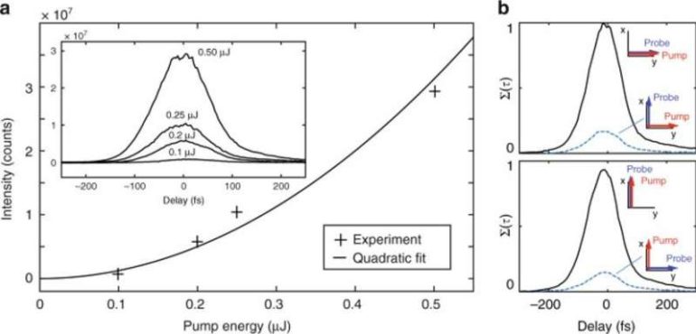
In this work, Xie and Meyer et al. showed how pump-induced micro-grating can be generated from the electronic Kerr effect—a phenomenon where the refractive index of a material changes due to an applied electric field—to provide a detailed in-situ characterization of a weak probe pulse. The scientists validated the measured diffracted signal and showed the validity of the measurement for all combinations of input pump and probe polarizations. They first reported on the validation of the technique, followed by the optimization of the probe pulse. Then they optimized the duration of the probe pulse to characterize both polarizations and showed how the method is very useful to detect spectral phase differences in the optical path of the pump and probe beams.


Spatial confinement of the synchronization
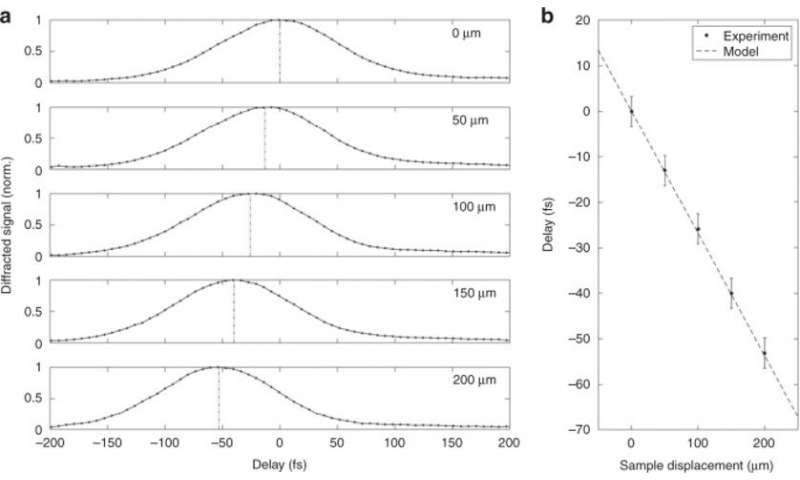
During the experiments, Xie and Meyer et al. defined the synchronization criterion of the pump and probe pulses for a precise location of focus in the sample and localized the interaction region between the pump and probe down to tens of micrometers. The strong localization of the experiment allowed them to retrieve the effect of the difference in group velocities on the pump-probe synchronization. The probe pulse can generate a pulse front tilt, which can limit ultrafast imaging experiments. To solve this, Xie and Meyer et al. used an aberration-free prism compressor by using two prisms that were perfectly parallel, although the parallelism can experimentally deviate by several milliradians. This deviation has a dramatic impact on the probe pulse. The team therefore used transient grating to offer a straightforward visualization of the pulse front tilt and then effectively resolved it by accurately adjusting the parallelism between the compressor prisms. The work showed excellent agreement between the experiments and simulations. The transient grating diagnostic introduced in this work was helpful to accurately remove pulse front tilt even for faint changes in the deviation angle of the prism compressor.
![Cross-correlation of pulses with angular and temporal dispersion. In the table, each trace shows the diffraction efficiency in arbitrary units as a function of delay (vertical axis) and spatial direction ky (horizontal axis, ky = [−1.03; 1.03] μm−1). The left table shows experimental results for 15 different combinations of temporal chirp ϕ2 and angular dispersion. The angular dispersion has been numerically characterized from the prism angle mismatch. The value of second order phase ϕ2 has been characterized from the prism insertions in the prism compressor (first row 3 mm, second row 2 mm, and last row 0 mm. The latter is the position for optimal pulse compression). For each trace, the horizontal axis scale has been converted to wavelength using the angular dispersion coefficient. When the angular dispersion is removed (central column), all wavelengths have the same direction ky. In this case, the lateral width of the spot is simply determined by the Gaussian beam size. To show the consistency of the results, the rightmost column show three cases (A, B, C) where analytical formula for the diffraction efficiency of the transient grating has been integrated using the parameters extracted from the ZEMAX simulations of the misaligned prism compressor. Light: Science & Applications, 10.1038/s41377-021-00562-1 In-situ diagnostic of femtosecond laser probe pulses for ultrafast imaging applications](https://techandsciencepost.com/wp-content/uploads/2021/07/1625582135_688_In-situ-diagnostic-of-femtosecond-laser-probe-pulses-for-ultrafast-imaging.jpg)
Outlook
In this way, Chen Xie, Remi Meyer and colleagues devised an extremely localized in-situ diagnostic method to allow the characterization and synchronization of a weak probe pulse with a higher intensity pump. The diagnostic is highly flexible to diverse pump-probe crossing geometries to characterize the probe pulse. The technique is also valid for a variety of pulse durations and is relevant even in the presence of spherical aberrations and widely applicable across most ultrafast imaging and pump-probe experiments. The results have diverse applications and can be useful to determine transient phenomena at the micron-scale as well as understand laser-matter interactions within condensed matter.
High-resolution, terahertz-driven atom probe tomography
More information:
Xie C., Meyer R. et al. In-situ diagnostic of femtosecond laser probe pulses for high resolution ultrafast imaging, Light: Science & Applications doi.org/10.1038/s41377-021-00562-1
Wrinkler T. et al. Laser amplification in excited dielectrics., Nature Physics, doi.org/10.1038/nphys4265
Frontiers in ultrashort pulse generation: pushing the limits in linear and nonlinear optics. Science, 10.1126/science.286.5444.1507
2021 Science X Network
Citation:
In-situ diagnostic of femtosecond laser probe pulses for ultrafast imaging applications (2021, July 6)
retrieved 6 July 2021
from https://phys.org/news/2021-07-in-situ-diagnostic-femtosecond-laser-probe.html
This document is subject to copyright. Apart from any fair dealing for the purpose of private study or research, no
part may be reproduced without the written permission. The content is provided for information purposes only.


