A microphysiological system (MPS), also known as an organ-on-a-chip, is a 3D organ construct using human cells that help reveal how organs respond to drugs and environmental stimuli.
Now, Tohoku University researchers have developed a new analytical method that visualizes cell functions in MPS using scanning probe microscopy (SPM).
SPM differs from optical microscopy since it employs fine probe scanning over a sample surface and then exploits the local interactions between the probe and the surface. The biggest advantage of SPM over conventional microscopy is that physical and chemical conditions can be acquired rapidly and as a high-resolution image.
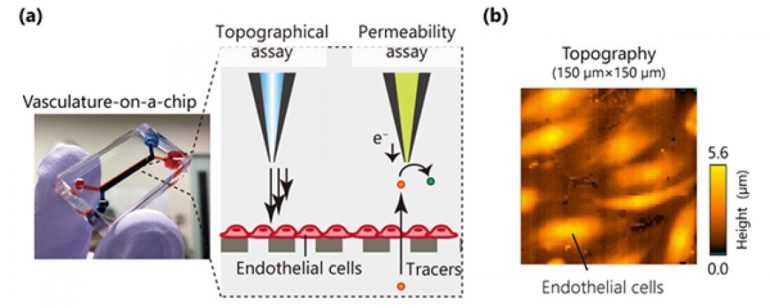
In this study, SPMs evaluated a vascular model (vasculature-on-a-chip) by scanning electrochemical microscopy (SECM) and scanning ion conductance microscopy (SICM). Using these SPMs, the researchers quantified the permeability and topographical information of the vasculature-on-a-chip.
“MPS shows potential to recapitulate the physiology and functions of their counterparts in the human body. Most research on this topic has focused on the construction of biomimetic organ models. Today, there is an increasing interest in developing sensing systems for MPS” said first author Yuji Nashimoto.
Some have touted electrochemical sensors to monitor MPS. However, most electrochemical sensors cannot acquire the spatial information of cell functions in MPS because they have only one sensor per one analyte. In contrast, SPM provides spatial information about cell functions rapidly.
“Our research group has developed various electrochemical imaging tools, SPMs and electrochemical arrays,” explained corresponding author Hitoshi Shiku.
“These devices will help usher in next-generation sensors in MPS.”
Imaging technique gives catalytic 2-D material engineering a better view

More information:
Yuji Nashimoto et al, Topography and Permeability Analyses of Vasculature‐on‐a‐Chip Using Scanning Probe Microscopies, Advanced Healthcare Materials (2021). DOI: 10.1002/adhm.202101186
Citation:
New imaging tool visualizes cell functions in a microphysiological system (2021, August 20)
retrieved 23 August 2021
from https://phys.org/news/2021-08-imaging-tool-visualizes-cell-functions.html
This document is subject to copyright. Apart from any fair dealing for the purpose of private study or research, no
part may be reproduced without the written permission. The content is provided for information purposes only.

