Texas A&M University researchers accomplished what was once considered impossible—they created a device capable of squeezing the quantum fluctuations of light down to a directed path and used it to enhance contrast imaging.
This one-of-a-kind “flashlight” was built to increase the signal-to-noise ratio present in Brillouin microscopy spectroscopic measurements that visually record the mechanical properties of structures inside living cells and tissues. Test results reveal the new source significantly increases image clarity and accuracy.
“This is a new avenue in research,” said Dr. Vladislav Yakovlev, University Professor in the Department of Biomedical Engineering in the College of Engineering. “We are specially designing light in such a way that it can improve contrast.”
“It’s a new milestone in the capabilities of Brillouin microscopy and imaging extensively used for bio systems,” said Dr. Girish Agarwal, University Distinguished Professor in the Department of Biological and Agricultural Engineering in the College of Agriculture and Life Sciences. “And it becomes part of an international effort to develop quantum sensors for diverse applications like brain imaging, biomolecule structure mapping and exploring underground oil and water sources by devising supersensitive gravimeters.”
A paper detailing the work was published in Optica.
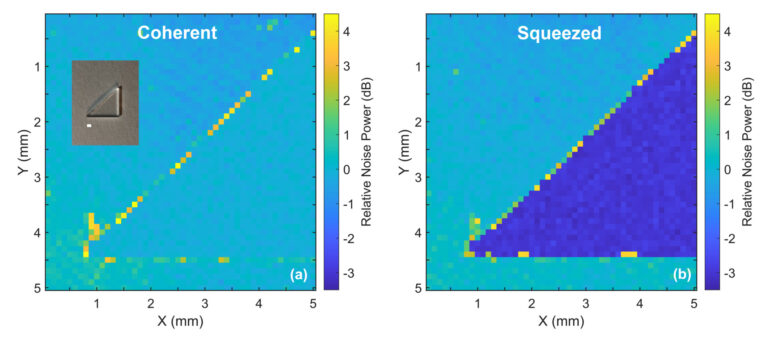
All instruments capable of capturing a picture or image also capture signal distortions, or noise, in the process. The distortions can stem from too much or too little light and even brightness or color issues from the environment around the subject. Most noise is unnoticed until the image is enlarged enough for the naked eye to see the unwanted pixels clearly.
Brillouin microscopy is the fundamental limit of reduced-scale measurement imaging currently possible. The process aims lasers at solid objects and measures the waves or signals of vibration made by the moving atoms and structures within the visibly unmoving material.
Noise produced at this scale can severely obscure the signals received, creating muddied images that are hard to interpret. Currently, all laser spectroscopy systems like Brillouin microscopy suffer from the natural and technical signal distortions associated with laser light, which is why newer light sources are needed.
Six years ago, Yakovlev attempted to improve the signal-to-noise ratio in Brillouin microscopy by using intense light sources. Unfortunately, overexposure to light damaged the cells he was imaging.
Yakovlev searched literature for answers and found a theory from the 1980s that postulated quantum light could solve the problem, though it didn’t mention how. Agarwal, an expert in quantum physics, came up with a possible way. Dr. Tian Li, then a postdoctoral researcher from the University of Maryland, was hired to create the first quantum light laboratory at Texas A&M. The laboratory space was provided by Dr. Marlan Scully, director of the Institute for Quantum Science and Engineering.
The team faced two significant challenges: finding funding for such a wild idea and finding graduate students and postdoctoral researchers to help them—ones who were willing to straddle the fields of biology and quantum physics.
After nearly two years of vigorous explorations, the device grew into a tabletop-sized contraption of complex optical configurations and measuring instruments that allowed the researchers to adjust, direct and efficiently manipulate and detect light. During that time, Li gained a better understanding of biology, and Yakovlev and Agarwal developed a mechanism to create the proper state and matter of light needed for noise reduction without damaging live cells.
Though the light-squeezing device can be adopted for other spectroscopic measurements like Raman scattering, Yakovlev and Agarwal are enhancing the capabilities of Brillouin microscopy to identify the viscous or elastic materials in biological systems. These systems control the physical properties of cells and cell structures and define everything from cell development to cancer progression.
Seeing details clearly makes a huge difference in biomedical breakthroughs.
“Each time you get a new telescope or something like gravitational-wave astronomy, you discover new things you can’t possibly see without it,” said Yakovlev. “The same thing works in biology. Before the invention of the microscope, we didn’t know that we consist of individual cells.”
So far, only the contrast of spectroscopy images has been improved, but Yakovlev and Agarwal are already working on Agarwal’s theory to enhance spatial resolution or the smallest details possible. And if the task leads to creating another complex device that pushes the limits of current technology, the researchers are ready and willing to make that happen.
“I love those types of projects where people tell you something will never work, and it works,” said Yakovlev. “I love challenges.”
More information:
Tian Li et al, Quantum-enhanced stimulated Brillouin scattering spectroscopy and imaging, Optica (2022). DOI: 10.1364/OPTICA.467635
Provided by
Texas A&M University College of Engineering
Citation:
Quantum light source advances bio-imaging clarity (2022, September 19)

