Researchers have developed a way to produce high-resolution virtual microscopy images that allow detailed visualization of tissue without time-consuming staining procedures. This approach is an important step toward the ability to perform histopathology analysis during surgery, rather than waiting days for results.
“In traditional histology, the cell nuclei and cytoplasm are differentiated by applying stains known as hematoxylin and eosin, commonly referred to as H&E,” said research team leader Roger J. Zemp from the University of Alberta in Canada. “Our virtual histology approach produces high-resolution nuclei and cytoplasm imaging with colorization that closely matches traditional H&E staining approaches.”
In the Optica Publishing Group journal Optics Letters, the researchers report the first label-free virtual H&E histology imaging with the resolution, speed and realism needed for performing diagnosis during a surgery. Histopathological analysis is often used to determine whether a tumor has been completely surgically removed. If the results show that cancer cells remain, the patient must go through another surgery to have those cells removed.
“We envision that virtual histopathology could be used on freshly resected tumor tissues within an intraoperative setting, providing direct feedback to surgeons,” said Zemp. “This could potentially reduce the need for repeat surgeries, improving outcomes for individual patients and reducing healthcare costs.”
Creating contrast
The new imaging method builds upon the research group’s previous work in which they successfully extracted cell nuclei information from tissue samples using ultraviolet light. However, replacing traditional histology with a virtual version requires a way to view both the cell nuclei and cytoplasm.
To accomplish this, the researchers combined pulsed ultraviolet and non-pulsed near-infrared light. The combination provides images that contain details about both the cell cytoplasm and the cell nuclei, making it possible to produce high-resolution virtual histology images without requiring any staining.
“The near-infrared reflectivity reveals information about the cell cytoplasm background,” said Nathaniel Haven, the paper’s first author. “When pulsed ultraviolet light is coupled into the system, it is absorbed by cell nuclei in a way that causes sharp changes in the near-infrared reflectivity and allows cell nuclei details to be extracted from the cell cytoplasm background. The ultraviolet beam can also be manipulated to provide reflectivity information that can be used to generate cytoplasm detail at much higher resolutions than is possible with just the near-infrared beam.”
However, using nanosecond-pulsed ultraviolet light to measure reflectivity at a single point on a sample is not easy with today’s image acquisition systems. To overcome this issue, the researchers used a custom peak detector circuit that holds the peak value of the reflectivity measurement so that it can be adequately sampled. The researchers also developed a colormap matching algorithm that applies realistic colors to the virtual images, making it intuitive for surgeons to interpret the images in real time.
Imaging breast cancer
Compared to what the researchers had previously accomplished, the new approach improves the resolution of cytoplasm images by a factor of five, which gives the cell nuclei and cytoplasm a uniform resolution in the virtual histology images.
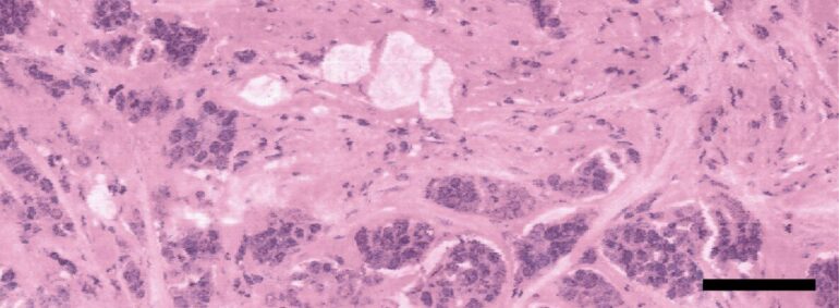
To demonstrate their technique, they compared virtual histology images of samples from human breast lumpectomy with traditional histology. The results showed that virtual H&E images of unstained tissues closely resembled true H&E-stained sections. They also assessed the preliminary diagnostic utility of the virtual approach by having a pathologist note diagnostic features of interest in the virtual images.

“The feedback we received from the pathologist was positive,” said co-author Matthew Martell. “If future validation studies are successful, we think that this approach could one day replace traditional histopathology in time-sensitive applications such as point-of-care biopsy analysis and cancer surgery.”
The researchers are now working toward conducting a single-blinded study that would statistically compare diagnoses based on their virtual histology approach with those using conventional histology. They also want to leverage artificial intelligence technology to rapidly interpret and flag areas for inspection, which would significantly speed up tissue examination.
More information:
Nathaniel J. M. Haven et al, Virtual histopathology with ultraviolet scattering and photoacoustic remote sensing microscopy, Optics Letters (2021). DOI: 10.1364/OL.436136
Provided by
The Optical Society
Citation:
Virtual histology approach lays groundwork for tissue analysis during surgery (2021, November 17)

