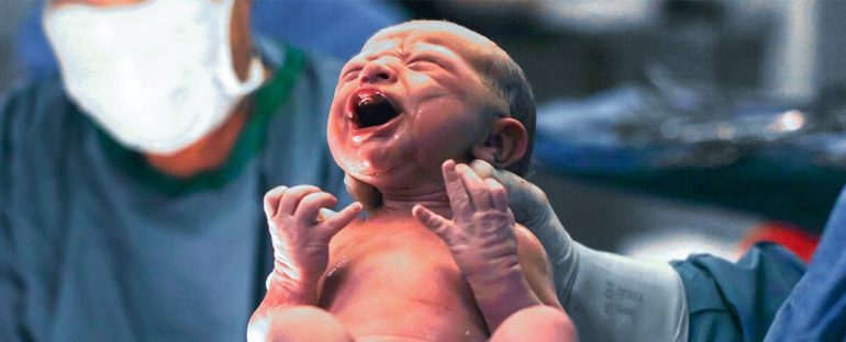
Nothing fills a parent with relief like the sound of their newborn’s cry. That first gulp of air defines such a critical moment in our lives, and yet strangely happens to be one of the least understood of any human behaviour.
Most of what we know about the physiological activity inside the lungs during a first breath comes either from pre-clinical studies – often involving animals – or through invasive observations that risk interfering with natural lung functions.
Now, using a delicate sensor-belt wrapped around the chests of full-term newborns moments after their delivery, a team of researchers from Australia, Germany, Switzerland, and Canada have recorded the changes in lung volume that occur in a baby’s first minutes outside the womb.
“Healthy term babies use remarkably complex methods of adapting to air-breathing at birth,” says clinical neonatologist David Tingay from the Murdoch Children’s Research Institute (MCRI) in Australia.
“There is a reason why parents, midwives, and obstetricians are pleased to hear those first life-affirming cries when a baby is born.”
The apparatus generated images of the babies’ chests, which together with video and audio data contributed to detailed descriptions of more than 1,400 breaths taken by 17 tiny humans all delivered through an elective caesarean section.
The results provided a statistical overview of the process of lung aeration in newborns – the physiological activity of preparing and inflating lungs to allow them to take over the placenta’s role of exchanging gases.
Putting it simply, while the babies are snug inside the uterus, they’re busy exercising their lungs: Though crammed full of a liquid secreted by the lung’s lining, they still move in a semblance of breathing to give the brain and muscles plenty of practice.
This training regime drops immediately before birth. The big squeeze that comes with being pushed through the vaginal canal forces out some of the liquid, while also providing a generous dose of adrenaline that tells the lungs to soak up as much of that fluid as they can.
That still leaves a fair bit of residue sloshing about, especially if delivery came via caesarean. Much of it is pushed back through the tissues with the first rush of air, with a soapy material called a surfactant helping the tiny air sacs shed their coating of fluid more easily and expand as wide as possible.
It’s little wonder that our first moments swanning about in the open air come with a gurgling cry.
Data from this new study help to flesh out the scene. That crying creates a unique pattern of gas flow that helps to preserve functional residual capacity; the air that we retain in our lungs after we breathe out.
This breathing wasn’t necessarily balanced between the two lungs either, tending to favour the right side more than the left.
Of all the breaths recorded, crying featured most in the first minute after birth as babies inflated their lungs; 6 minutes later, more than half of the babies’ breaths had eased into a regular flow called tidal breathing.
Having a signature of breathing patterns in healthy babies delivered via caesarean sets a foundation for exploring how lung aeration differs in other cases, such as in preterm infants.
Around 1 in 10 newborns, and virtually all children born prior to 37 weeks, require assistance in catching that first important breath.
For those who find this kind of research absolutely fascinating, MCRI have put together a detailed, but still easily accessible video clip explaining the study, which you can watch below.
“Improving interventions in the delivery room first requires understanding the processes that define success and failure of breathing at birth,” says Tingay.
In emergency cases this could require traumatic interventions such as intubation, which – although it saves lives – can damage airways and leave infants at risk of damage to airway tissues, infection, and respiratory illnesses.
Knowing when drastic actions need to be taken, and when they don’t, could help avoid putting newborns at unnecessary risk.
The equipment used in the study is about as non-invasive as it gets, so could be applied to most births, caesarean and vaginal, with virtually no risk to the newborn.
“This new technology not only allows us to see deep into the lungs but is also the only method we have of continuously imaging the lungs without using radiation or interrupting life-saving care,” says Tingay.
“This study has shown that babies’ lungs are far more complicated than traditional monitoring methods had previously suggested.”
This research was published in the American Journal of Respiratory and Critical Care Medicine.


