The microscope is an iconic symbol of the life sciences – and for good reason. From the discovery of the existence of cells to the structure of DNA, microscopy has been a quintessential tool of the field, unlocking new dimensions of the living world not only for scientists but also for the general public.
For the life sciences, where understanding the function of a living thing often requires interpreting its form, imaging is vital to confirming theories and revealing what is yet unknown.
This selection of stories from The Conversation’s archive presents a few ways in which microscopy has contributed to different forms of scientific knowledge, including techniques that take visualization beyond sight altogether.
1. Seeing as identifying
Over the past few centuries, the microscope has undergone a gradual but significant evolution. Each advance has allowed researchers to see increasingly smaller and more fragile structures and biomolecules at increasingly higher resolution – from cells, to the structures within cells, to the structures within the structures within cells, down to atoms.
But there is still a resolution gap between the smallest and largest structures of the cell. Biophysicist Jeremy Berg drew an analogy to Google Maps: Though scientists could see the city as a whole and individual houses, they couldn’t make out the neighborhoods.
“Seeing these neighborhood-level details is essential to being able to understand how individual components work together in the environment of a cell,” he writes.
Scientists are working to bridge that resolution gap. Improvements to the 2014 Nobel Prize-winning superresolution microscopy, for example, have enhanced the study of lengthy processes like cell division by capturing images across a range of size and time scales simultaneously, bringing clarity to details traditional microscopes tend to blur.
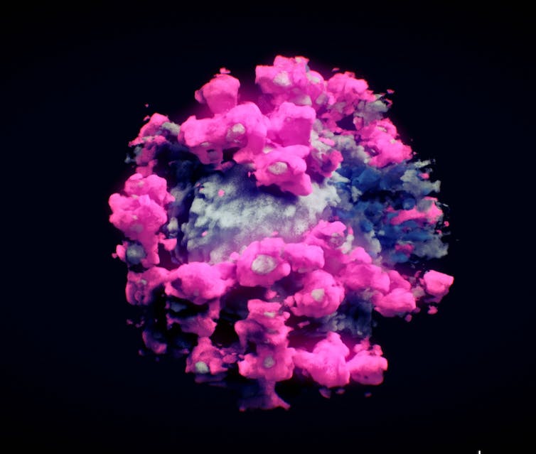
Cryo-electron tomography shows what molecules look like in high resolution – in this case, the virus that causes COVID-19.
Nanographics, CC BY-SA
Another technique, cryo-electron microscopy, or cryo-EM, won a Nobel Prize in 2017 for bringing even more complex, dynamic molecules into view by flash-freezing them. This creates a protective glasslike shell around samples as they’re bombarded by a beam of electrons to create their photo op. Cryo-ET, a specialized type of cryo-EM, can construct 3D images of molecular structures within their natural environments.
These techniques not only generate images at or near atomic resolution but also preserve the natural shape of difficult-to-capture biomolecules of interest. Researchers were able to use cryo-EM, for instance, to capture the elusive structure of the protein on the surface of the shape-shifting hepatitis C virus, providing key information for a future vaccine.
Further enhancements to science’s visual acuity will reveal more of the fine details of the building blocks of life.
“I…

