
Like the people they make up, cells communicate by bumping into one another and exchanging handshakes. Unlike people, cells perform these handshakes using the diverse range of sugar molecules coating their surface like trees covering a landscape. Handshakes between these sugar molecules, or glycans, trigger cells to react in specific ways toward each other, such as escape, ignore or destroy.
Figuring out the “body language” of glycans during these handshakes can provide clues to how cancers, infections and immune systems work, as well as solutions to health and sustainability challenges society faces today.
What are glycans?
Each glycan molecule is made up of a network of individual sugar molecules bonded together. The vast number of possible glycan structures that can be built from connecting these sugar molecules together allows glycans to store rich information.
Because all living cells are covered with sugars, glycans act like ID cards for cells. They display the cell’s identity, such as whether it’s a bacteria or human cell, and its state, such as whether it’s healthy or cancer, to the rest of the body and allow other cells to recognize and respond to it. For example, these identifying signs allow our immune cells to recognize and clear out harmful bacteria and cancerous cells while leaving healthy cells in peace.
An example of how glycan-stored information is important to daily life is your blood type. Glycans are chemically bonded to proteins and lipids on the surface of red blood cells. Notably, the surface of type A red blood cells have glycans that differ from the glycans on the surface of type B and type O red blood cells. Knowing what blood type you have is important to avoid an unwanted immune response during blood transfusions.
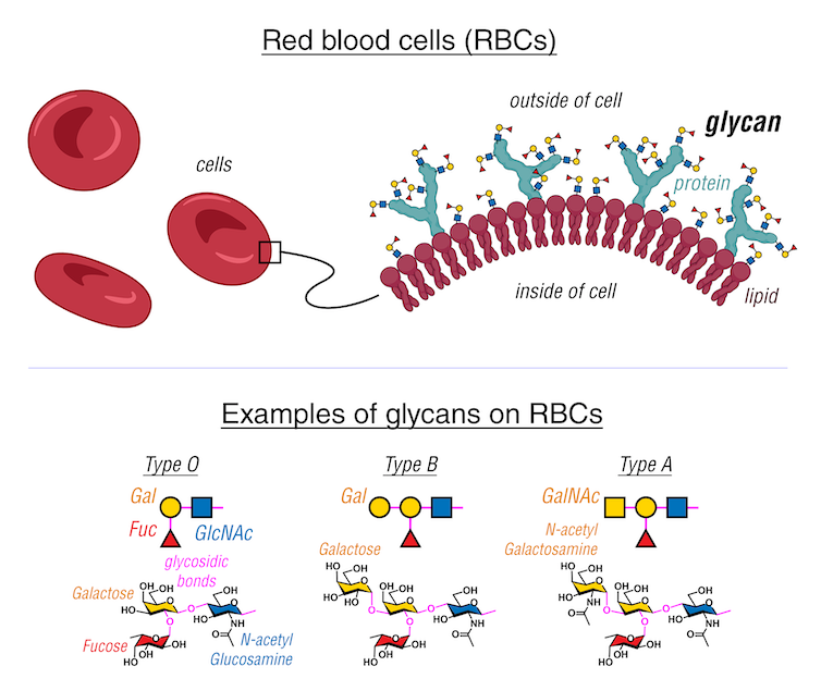
Your blood type is determined by the types of glycans, depicted here in circles and triangles, on your red blood cells.
Kelvin Anggara/Created with BioRender.com, CC BY-SA
Proteins decorated with glycans, or glycoproteins, and lipids decorated with glycans, or glycolipids, are ubiquitous in nature.
For example, distinctive glycoproteins cover the surface of the viruses that cause COVID-19, HIV and H1N1 influenza and help them infect cells. Glycolipids also coat many bacteria, allowing them to stick to their hosts and protect them from viruses and immune cells.
More recently, researchers discovered pieces of genetic material decorated with glycans on the surfaces of mammalian cells, challenging the long-standing notion that genetic material could be found only in the nucleus of cells and launching research to determine the functions of these glycans. One recent study showed that these molecules are vital in attracting immune cells toward infected or injured tissues.
How do cells read glycans?
In addition to the rich biological information contained in glycans, their easily accessible locations on cell surfaces make them highly attractive…

