Using a new model of brain activity, Indiana University computational neuroscientists Maria Pope, Richard Betzel and Olaf Sporns are exploring striking bursts of activity in the human brain that have not been examined before. These bursts may have potential to serve as biomarkers for brain disease and conditions such as depression, schizophrenia, dementia, and ADHD.
While analyzing human neuroimaging data, the IU research team discovered short bursts of activity that form ongoing “events” in the brain and are always taking place no matter the activity or state of the brain. In the course of a 10-minute brain scan, these events will occur roughly 10 to 20 times, each lasting for just a few seconds, the researchers found.
“What people had not seen is that how brain regions talk to each other is punctuated by these brief moments that are just a few seconds long during which there’s a lot happening,” said Olaf Sporns, who is Distinguished Professor and Robert H. Shaffer Chair in the College of Arts and Sciences Department of Psychological and Brain Sciences at IU Bloomington.
“Now that we see them, we’ve focused on those moments to get a picture of how specific brain regions link up and talk to each other during these events.”
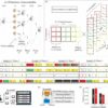
To begin investigating the workings of these mysterious events, the team built a computational model. Led by Maria Pope, a graduate student in Sporns’ lab and a dual Ph.D. candidate in neuroscience and informatics, the group used neuroimaging data of a human brain to build a model replicating its connections. The model was then simulated in a state similar to the resting brain to create synthetic MRI signals, using mathematical equations that reenact neuronal activity.
The model showed burst-like events just like those seen in human brain recordings.
The paper outlining the model and describing how it compares to the real brain was published in the November 16 issue of the Proceedings of the National Academy of Sciences.
“The model shows us that these events are guided by the brain’s structural network,” Pope said. “They are tied to the physical structure of brain.”
More specifically, the events originate in clusters of neurons and brain regions that are densely interconnected and momentarily light up together. Sporns compared the pattern to an orchestra playing a piece of music.
“There are moments when the orchestra comes together and there’s a theme. They are not just playing a single note for 10 minutes. There are brief moments in which coordinated activity dominates and at other times there might be much less,” Sporns said. “This ebb and flow of coordination is something we also see in the brain, and our model can reproduce it. Clusters of brain regions combine in different ways. It’s not just one pattern, but multiple variations on a theme.”
The new model’s outcome, Sporns suggested, is a potential game changer.
“Functional connectivity has been a strong focus in research as a potential biomarker for brain disorders and has been related to conditions such as depression, schizophrenia, dementia, and ADHD. And researchers have tried for years to use brain simulations in clinical applications for modeling lesions or diseases,” Sporns said. “This new model gives us a better lens through which to look at the brain, to see more clearly what goes on under both normal and abnormal conditions.”
The researchers are now delving further into why the human brain employs these brief bursts of activity.
“Perhaps the brain has developed this type of activity because it’s beneficial. Something about the structure of events may be useful to the brain,” Pope said. “For example, many kinds of networked systems have to do occasional system updates or resets, taking some kind of globally useful information and communicating it to the rest of the system.”
Answers to these questions may have implications not only for understanding the brain, but also for the study of neural networks and artificial intelligence.
“A clearer mapping of structure and function at the individual level could have implications for how we diagnose neurological disease and lead to personalized treatment and intervention,” said Betzel, professor in the College of Arts and Sciences Department of Psychological and Brain Sciences.
The study was funded by National Science Foundation grant “NRT: Interdisciplinary Training in Complex Networks and Systems” (NSF 1735095) to Maria Pope and National Science Foundation grant “NCS-FO: Edge-centric maps of functional brain network organization and dynamics” (NSF 2023985) to Richard Betzel and Olaf Sporns. Other co-authors include Makoto Fukushima, Division of Information Science, Graduate School of Science and Technology, and the Data Science Center, Nara Institute of Science and Technology, Ikoma, Nara, Japan and Center for Information and Neural Networks, National Institute for Information and Communications Technology, Suita, Osaka, Japan.


