The loss and return of consciousness is linked to the same network of brain regions for both sleep and anesthesia, according to new research published in JNeurosci.
The biological basis of consciousness has confounded scientists for centuries. Our experimental techniques falter, as the effects of sleep and anesthetic drugs alter brain activity beyond changes in consciousness. In addition, behavior does not always reveal someone’s state of consciousness. An unresponsive person might still be aware of their surroundings (connected), or unaware but still experiencing their internal world (disconnected).
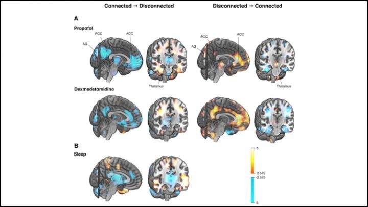
Scheinin et al. sought networks associated with human consciousness by measuring the brain activity of adult males with PET as they fell asleep and went under anesthesia. The research team woke participants mid-experiment to interview them and confirm their state of connectedness. Changes in connectedness corresponded to the activity of a network comprised of regions deep inside the brain: the thalamus, anterior and posterior cingulate cortex, and angular gyri. These regions exhibited less blood flow when a participant lost connectedness and more blood flow when they regained it. The pattern held true for both sleep and anesthesia, indicating the changes corresponded to connectedness rather than the effects of sleep or drugs, and that the network may be imperative for human consciousness.
Brain study suggests consciousness a matter of optimal degree of connectedness in neural network
More information:
Foundations of Human Consciousness: Imaging the Twilight Zone,JNeurosci (2020). DOI: 10.1523/JNEUROSCI.0775-20.2020
Provided by
Society for Neuroscience

Citation:
The brain network driving changes in consciousness (2020, December 28)
retrieved 28 December 2020
from https://medicalxpress.com/news/2020-12-brain-network-consciousness.html
This document is subject to copyright. Apart from any fair dealing for the purpose of private study or research, no
part may be reproduced without the written permission. The content is provided for information purposes only.

