All memory storage devices, from your brain to the RAM in your computer, store information by changing their physical qualities. Over 130 years ago, pioneering neuroscientist Santiago Ramón y Cajal first suggested that the brain stores information by rearranging the connections, or synapses, between neurons.
Since then, neuroscientists have attempted to understand the physical changes associated with memory formation. But visualizing and mapping synapses is challenging to do. For one, synapses are very small and tightly packed together. They’re roughly 10 billion times smaller than the smallest object a standard clinical MRI can visualize. Furthermore, there are approximately 1 billion synapses in the mouse brains researchers often use to study brain function, and they’re all the same opaque to translucent color as the tissue surrounding them.
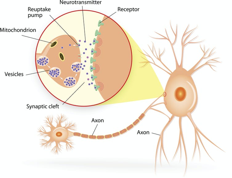
Synapses comprise the very end of the transmitting neuron, the very beginning of the receiving neuron, and the tiny gap between them.
ttsz/iStock via Getty Images Plus
A new imaging technique my colleagues and I developed, however, has allowed us to map synapses during memory formation. We found that the process of forming new memories changes how brain cells are connected to one another. While some areas of the brain create more connections, others lose them.
Mapping new memories in fish
Previously, researchers focused on recording the electrical signals produced by neurons. While these studies have confirmed that neurons change their response to particular stimuli after a memory is formed, they couldn’t pinpoint what drives those changes.
To study how the brain physically changes when it forms a new memory, we created 3D maps of the synapses of zebrafish before and after memory formation. We chose zebrafish as our test subjects because they are large enough to have brains that function like those of people, but small and transparent enough to offer a window into the living brain.
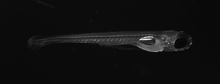
Zebrafish are particularly fitting models for neuroscience research.
Zhuowei Du and Don B. Arnold, CC BY-NC-ND
To induce a new memory in the fish, we used a type of learning process called classical conditioning. This involves exposing an animal to two different types of stimuli simultaneously: a neutral one that doesn’t provoke a reaction and an unpleasant one that the animal tries to avoid. When these two stimuli are paired together enough times, the animal responds to the neutral stimulus as if it were the unpleasant stimulus, indicating that it has made an associative memory tying these stimuli together.
As an unpleasant stimulus, we gently heated the fish’s head with an infrared laser. When the fish flicked its tail, we took that as an indication that it wanted to escape. When the fish is then exposed to a neutral stimulus, a light turning on, tail flicking meant that it’s recalling what happened when it previously…

