Scientists at the University of Nottingham have developed an ultrasonic imaging system, which can be deployed on the tip of a hair-thin optical fiber, and will be insertable into the human body to visualize cell abnormalities in 3D.
The new technology produces microscopic and nanoscopic resolution images that will one day help clinicians to examine cells inhabiting hard-to-reach parts of the body, such as the gastrointestinal tract, and offer more effective diagnoses for diseases ranging from gastric cancer to bacterial meningitis.
The highlevel of performance the technologydeliversis currentlyonly possible instate-of-the-artresearch labswith large, scientific instruments—whereasthis compact system has the potential to bring it into clinical settings to improve patient care.
The Engineering and Physical Sciences Research Council (EPSRC)-funded innovation also reduces the need for conventional fluorescent labels—chemicals used to examine cell biology under a microscope—which can be harmful to human cells in large doses.
The findings are being reported in a new paper, titled “Phonon imaging in 3D with a fiber probe,” published in Light: Science & Applications.
Paper author Salvatore La Cavera, an EPSRC Doctoral Prize Fellow from the University of Nottingham Optics and Photonics Research Group, said of the ultrasonic imaging system: “We believe its ability to measure the stiffness of a specimen, its bio-compatibility, and its endoscopic-potential, all while accessing the nanoscale, are what set it apart. These features set the technology up for future measurements inside the body; towards the ultimate goal of minimally invasive point-of-care diagnostics.”
Currently at prototype stage, the non-invasive imaging tool, described by the researchers as a “phonon probe,” is capable of being inserted into a standard optical endoscope, which is a thin tube with a powerful light and camera at the end that is navigated into the body to find, analyze, and operate on cancerous lesions, among many other diseases. Combining optical and phonon technologies could be advantageous; speeding up the clinical workflow process and reducing the number of invasive test procedures for patients.
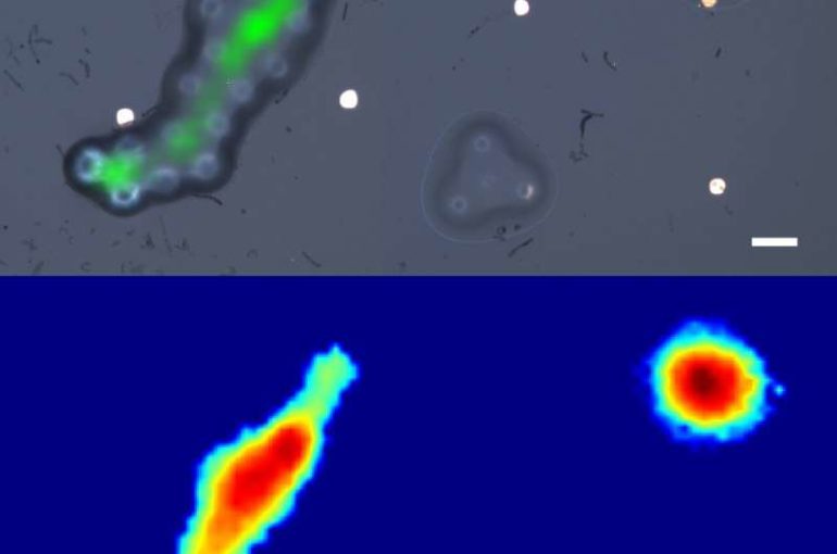
3D mapping capabilities
Just as a physician might conduct a physical examination to feel for abnormal ‘stiffness’ in tissue under the skin that could indicate tumors, the phonon probe will take this ‘3D mapping’ concept to a cellular level.
By scanning the ultrasonic probe in space, it can reproduce a three-dimensional map of stiffness and spatial features of microscopic structures at, and below, the surface of a specimen (e.g. tissue); it does this with the power to image small objects like a large-scale microscope, and the contrast to differentiate objects like an ultrasonic probe.
“Techniques capable of measuring if a tumor cell is stiff have been realized with laboratory microscopes, but these powerful tools are cumbersome, immobile, and unadaptable to patient-facing clinical settings. Nanoscale ultrasonic technology in an endoscopic capacity is poised to make that leap,” adds Salvatore La Cavera.
How it works
The new ultrasonic imaging system uses two lasers that emit short pulses of energy to stimulate and detect vibrations in a specimen. One of the laser pulses is absorbed by a layer of metal—a nano-transducer (which works by converting energy from one form to another) – fabricated on the tip of the fiber; a process which results in high-frequency phonons (sound particles) getting pumped into the specimen. Then a second laser pulse collides with the sound waves, a process known as Brillouin scattering. By detecting these “collided” laser pulses, the shape of the traveling sound wave can be recreated and displayed visually.
The detected sound wave encodes information about the stiffness of a material, and even its geometry. The Nottingham team was the first to demonstrate this dual-capability using pulsed lasers and optical fibers.
The power of an imaging device is typically measured by the smallest object that can be seen by the system, i.e. the resolution. In two dimensions the phonon probe can “resolve” objects on the order of 1 micrometer, similar to a microscope; but in the third dimension (height) it provides measurements on the scale of nanometres, which is unprecedented for a fiber-optic imaging system.

Future applications
In the paper, the researchers demonstrate that the technology is compatible with both a single optical fiber and the 10,000 to 20,000 fibers of an imaging bundle (1mm in diameter), as used in conventional endoscopes.

Consequently, superior spatial resolution and wide fields of view could routinely be achieved by collecting stiffness and spatial information from multiple different points on a sample, without needing to move the device—bringing a new class of phonon endoscopes within reach.
Beyond clinical healthcare, fields such as precision manufacturing and metrology could use this high-resolution tool for surface inspections and material characterisation; a complementary or replacement measurement for existing scientific instruments. Burgeoning technologies such as 3D bio-printing and tissue engineering could also use the phonon probe as an inline inspection tool by integrating it directly to the outer diameter of the print-needle.
Next, the team will be developing a series of biological cell and tissue imaging applications in collaboration with the Nottingham Digestive Diseases Center and the Institute of Biophysics, Imaging and Optical Science at the University of Nottingham; with the aim to create a viable clinical tool in the coming years.
Using holographic endoscopes to observe distant objects
More information:
Salvatore La Cavera et al. Phonon imaging in 3D with a fibre probe, Light: Science & Applications (2021). DOI: 10.1038/s41377-021-00532-7
Provided by
University of Nottingham
Citation:
World’s first fiber-optic ultrasonic imaging probe for future nanoscale disease diagnostics (2021, April 30)
retrieved 3 May 2021
from https://phys.org/news/2021-04-world-fiber-optic-ultrasonic-imaging-probe.html
This document is subject to copyright. Apart from any fair dealing for the purpose of private study or research, no
part may be reproduced without the written permission. The content is provided for information purposes only.

